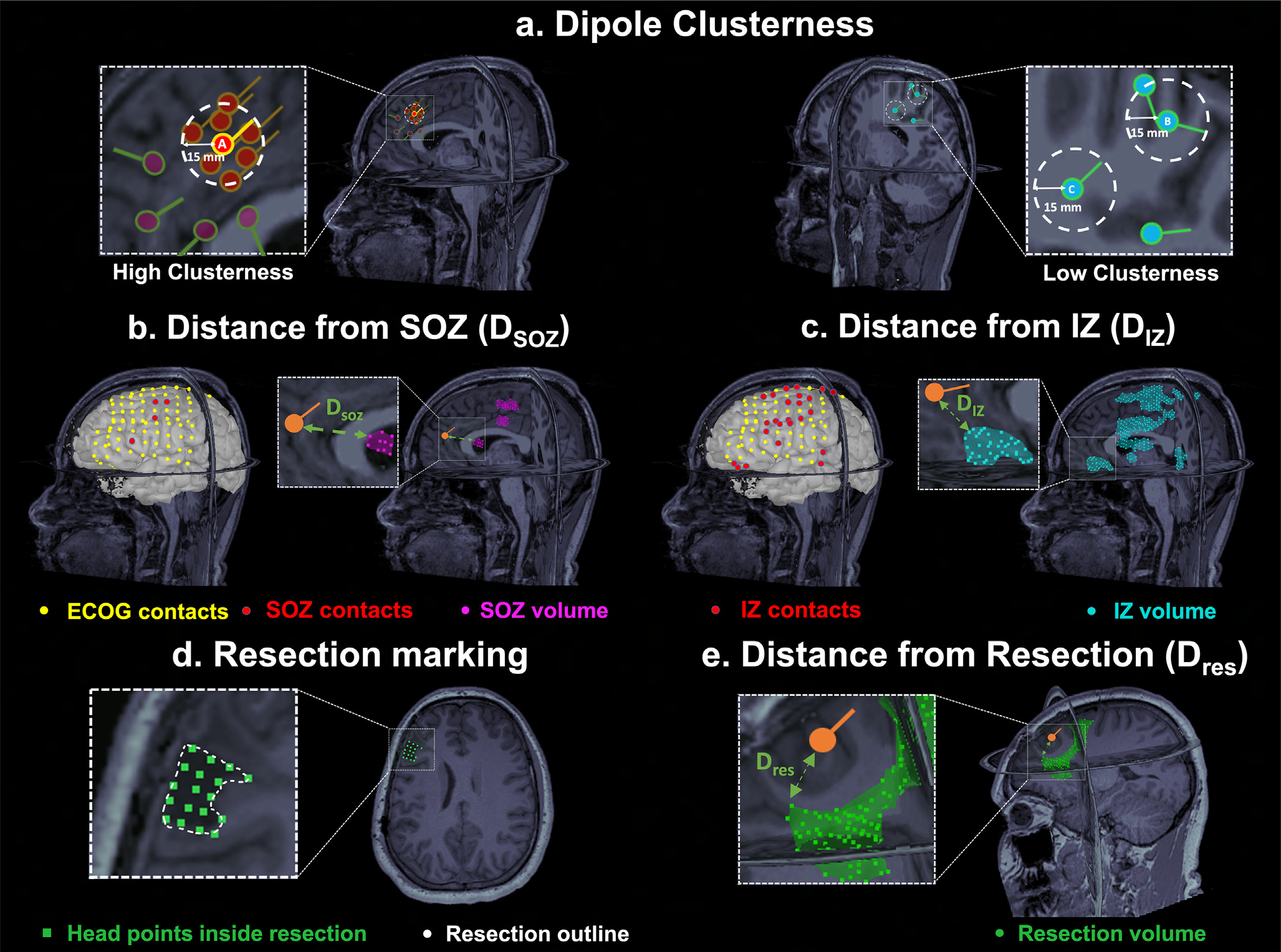
Fig. 1. Estimation of Dipole Clusterness and Distances from Seizure Onset Zone (DSOZ). Irritative Zone (DIZ), and Resection (Dres).

(a) Left: Example of a dipole (dipole A) with high clusterness, i.e. high number of surrounding dipoles (n = 7) within a radius of 15 mm (white dash circle). Dipoles which are outside the 15-mm radius of dipole A do not contribute to clusterness. Right: Example of dipoles with low clusterness (i.e. with one or two surrounding dipoles). (b) Intracranial EEG (icEEG) contacts belonging to the Seizure Onset Zone (SOZ) (in red) defined the SOZ volume (purple) in Magnetic Resonace Imaging (MRI) space. Euclidian distance (green arrow) of each dipole (orange) from the closest point of the SOZ volume was computed (DSOZ). (c) icEEG contacts belonging to the Irritative Zone (IZ) (in red) defined the IZ volume (cyan). Euclidian distance (green arrow) of each dipole from the closest point of the IZ volume was computed (DIZ). (d) Definition of the resected cavity (green points) on the postoperative MRI after its coregistration with the preoperative MRI. (e) Euclidian distance (green arrow) of a dipole (orange) from the closest point of the resection (DRES).
