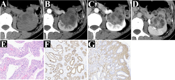
FIGURE 4.

A 49‐year‐old woman with flank pain (case 15). A cystic‐solid mass is shown on an axial enhancement multidetector computed tomography (MDCT) (a), which displayed slight enhancement with multi‐septate cystic and solid components on the corticomedullary phase (b), prolonged enhancement on the nephrographic phase (c) and the excretory phase (d), and was confirmed as mixed epithelial and stromal tumor of the kidney (MESTK) by histopathological (e) and immunohistochemical ((f) the positive for pan‐cytokeratin in the epithelium; (g) positive for vimentin in the stromal area). The patient (case 15) was classified into cystic type (IV category). Compared with Figure 3 (case 13), it has a variable proportion of cystic and solid components
