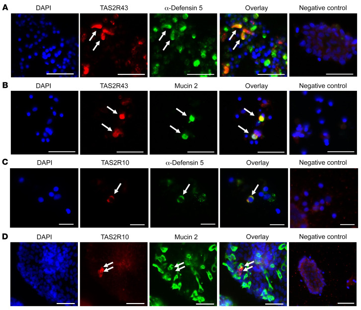
Figure 1. Colocalization of TAS2R43 and TAS2R10 with Paneth or goblet cells in primary crypts from patients with obesity.
Representative double-immunofluorescence images of jejunal crypts from obese patients. Crypts were stained for TAS2R43 (A and B) or TAS2R10 (C and D) (red) and α-defensin 5 (in Paneth cells) or mucin 2 (in goblet cells) (green). Images in A–C show colocalization, whereas the images in D show a TAS2R10+ cell in close proximity to a goblet cell, but no colocalization. Nuclei were stained with DAPI (blue). Scale bars: 20 μm (C) and 50 μm (A, B, and D). Each colocalization study was repeated in crypts derived from at least 3 patients with obesity.

