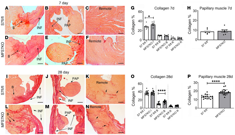
Figure 4. Myofibroblast-specific Smad7 loss accentuates postinfarction fibrosis in the border zone and in the papillary muscles.
Collagen staining was performed using picrosirius red, and the collagen-stained area was assessed in the papillary muscle (PAP), infarcted (INF), border (B), and remote remodeling areas of Smad7fl/fl (S7 fl/fl) and MFS7KO hearts at 7 (A–F) and 28 days (I–N) of coronary occlusion. (G and H) Quantitative analysis demonstrates increased collagen deposition in the infarct zone of MFS7KO hearts compared with Smad7fl/fl at 7 days after infarction. (O and P) Twenty-eight days after infarction, increased collagen deposition is noted in the infarct border zone (L) and in the papillary muscle (M) of MFS7KO hearts compared with Smad7fl/fl, with comparable collagen levels in the infarct zone and the remote myocardium. For comparisons between multiple groups (G and O), 1-way ANOVA was performed followed by Tukey’s multiple comparison test. For comparisons between 2 groups (H and P), unpaired 2-tailed Student’s t test with Welch’s correction for unequal variances was performed (7 days Smad7fl/fl, n = 6; MFS7KO, n = 7; 28 days Smad7fl/fl, n = 18; MFS7KO, n = 21). *P < 0.05; ****P < 0.0001. Scale bars: 100 μm.

