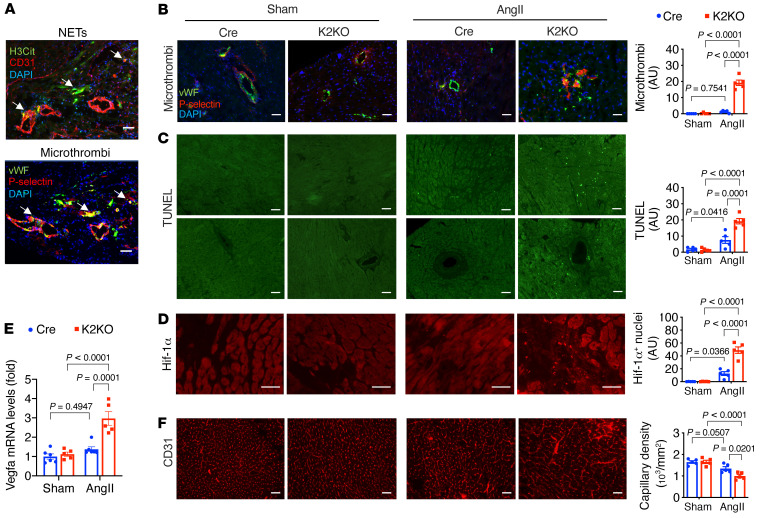
Figure 5. AngII-induced NET formation triggers microthrombosis and myocardial injury.
(A and B) Immunostaining of H3Cit, vWF, P-selectin, and CD31. (C) TUNEL staining to assess cell death. Upper: Intramuscular regions. Lower: Perivascular regions. (D) Immunostaining of HIF1α protein. HIF1α-positive nuclei were counted. (E) Myocardial expression of Vegfa mRNA. (F) Myocardial capillary density assessed by CD31 immunostaining. AngII infusion: 1 week (A–D) or 4 weeks (E and F). P values are from 2-way ANOVA with Tukey’s correction (B–F). Representative images from 5 to 6 mice in each group. Scale bars: 25 μm.

