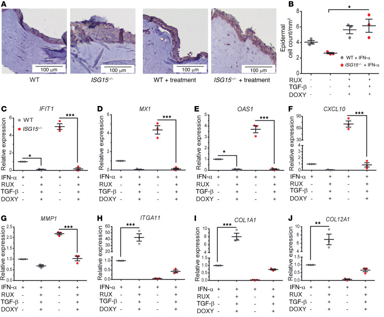
Figure 11. Combined treatment with RUX, DOXY, and TGF-β1 normalizes epidermis formation, inflammation, and collagen synthesis in the ISG15–/– 3D model.
3D epidermis models were generated as in Figure 7 and cultured in the presence of RUX (0.5 μM), DOXY (50 μM), and TGF-β (10 nM) from day 8 to day 14. (A) H&E stains showing thicker, less compact epithelial layer in the ISG15–/– model, which appears to normalize under treatment. Scale bars: 100 μm. (B) Triple treatment leads to increase of epithelial cell density of the ISG15–/– model to WT density. (C–J) Triple treatment reduces expression of IFN-regulated mRNAs (C–F) and MMP1 (G), and increases expression of ITGA11 (H), COL1A1 (I), and COL12A1 (J). n = 3, ± SEM; *P < 0.05, **P < 0.01, ***P < 0.001 (Student’s t test).

