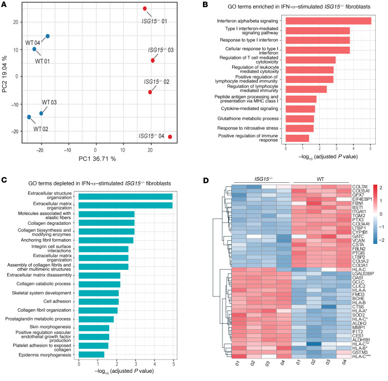
Figure 3. Proteomic analysis reveals major dysregulation in connective tissue homeostasis in ISG15–/– fibroblasts after IFN-α stimulation.
Immortalized dermal fibroblasts carrying either the native ISG15 locus (WT) or a naturally occurring loss-of-function mutation (ISG15–/–) were stimulated with IFN-α (1000 IU/mL) for 24 hours and subjected to global proteome analysis (n = 4). (A) Principal component analysis demonstrating clear separation between WT and ISG15–/– cells based on their protein expression patterns. (B and C) Enrichment analysis based on the proteome data used as input for A. GO terms relating to IFN responses, other inflammatory processes, and stress responses are enriched in ISG15–/– cells (B), whereas GO terms relating to epidermal and connective tissue/extracellular matrix homeostasis are depleted in ISG15–/– cells (C). X axes show Benjamini-Hochberg adjusted P values (–log10) for enrichment/depletion; only GO terms with adjusted P less than 0.05 are shown. (D) Hierarchical clustering of differentially abundant (adjusted P < 0.05) proteins showing upregulation of proinflammatory proteins, HLA molecules, and MMP1 but downregulation of collagen constituents and ROS scavengers in ISG15–/– cells.

