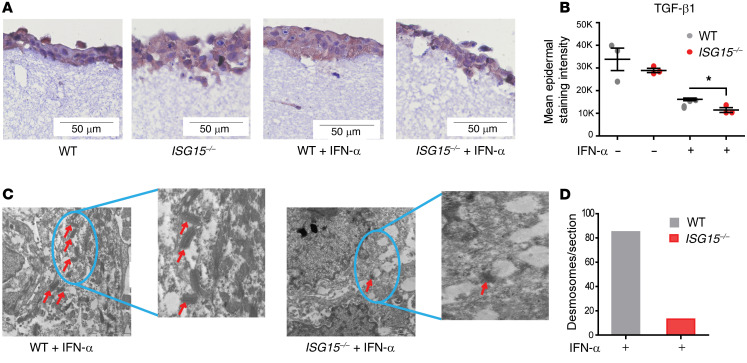
Figure 7. ISG15 deficiency causes histological disorganization and loss of desmosomes in a 3D epidermis model.
An epidermis-like structure was formed using coculture of HaCaT cells and immortalized dermal fibroblasts (both WT or both ISG15–/–) on a collagen matrix and analyzed by light and electron microscopy, RT-qPCR (ΔΔCt method, using unstimulated WT models as reference), and enzyme-linked immunoassay. (A) A compact epidermis forms in WT cells with and without IFN-α stimulation, whereas a loose arrangement is apparent when ISG15–/– cells are used. Visualization of the epidermis is enhanced by IHC staining for TGF-β1 (brown). Scale bars: 50 μm. (B) Lower TGF-β1 staining intensity in the epidermal layer of the ISG15–/– 3D model (based on automated image analysis of TGF-β1 IHC). A representative example comparing staining with specific and nonspecific (isotype control) antibody is shown in Supplemental Figure 7. (C and D) Desmosomes (identified by red arrows) are easily identified by transmission electron microscopy in the WT, but not in the ISG15–/– 3D model. Scale bars: 2 μm. (D) Markedly lower desmosome density in the ISG15–/– 3D model after IFN-α stimulation, as determined by 2 independent examiners by manual counting of typical structures in electron microscopic images.

