Figure 1.

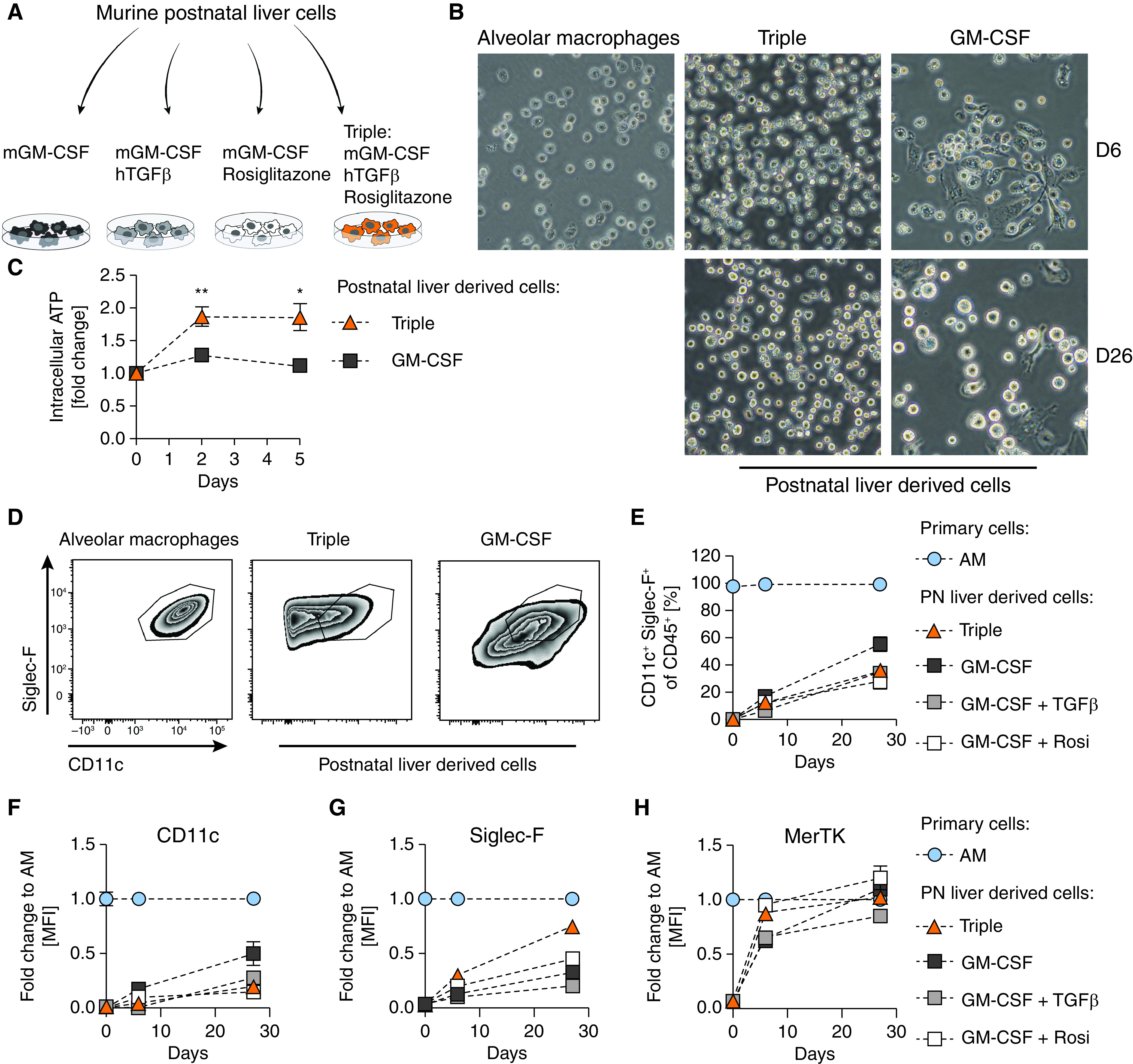
Murine AM-like cells can be derived from postnatal liver cells. (A) Experimental setup. (B) Primary AMs after 3 hours in culture and postnatal liver cells treated with murine GM-CSF (30 ng/ml) or murine GM-CSF (30 ng/ml) + human TGFβ (10 ng/ml) + rosiglitazone (1 µM) (Triple) after 6 days (D6) and 26 days (D26) in culture; 40 × magnification. (C) Cell proliferation of postnatal liver cells under indicated conditions over 5 days compared with time of seeding. (D) FACS analysis of Siglec-F and CD11c expression on primary AMs and postnatal liver cells grown under indicated conditions on D26. (E) Percentage of CD11c+Siglec-F+ cells in postnatal liver cell cultures at day of seeding, D6, and D26. (F) CD11c, (G) Siglec-F, and (H) MerTK mean fluorescence intensity degrees of postnatal liver cell cultures as fold change to primary AMs at indicated days. (D–H) Pregated on single, viable CD45+ cells. Graphs show means ± SEM of 3–4 biological replicates. Data are representative of at least two independent experiments. *P < 0.05 and **P < 0.01 (Student’s t test). AM = alveolar macrophages; GM-CSF = granulocyte-macrophage colony-stimulating factor; MFI = mean fluorescence intensity; PN = postnatal; Rosi = rosiglitazone; TGFβ = transforming growth factor β.
