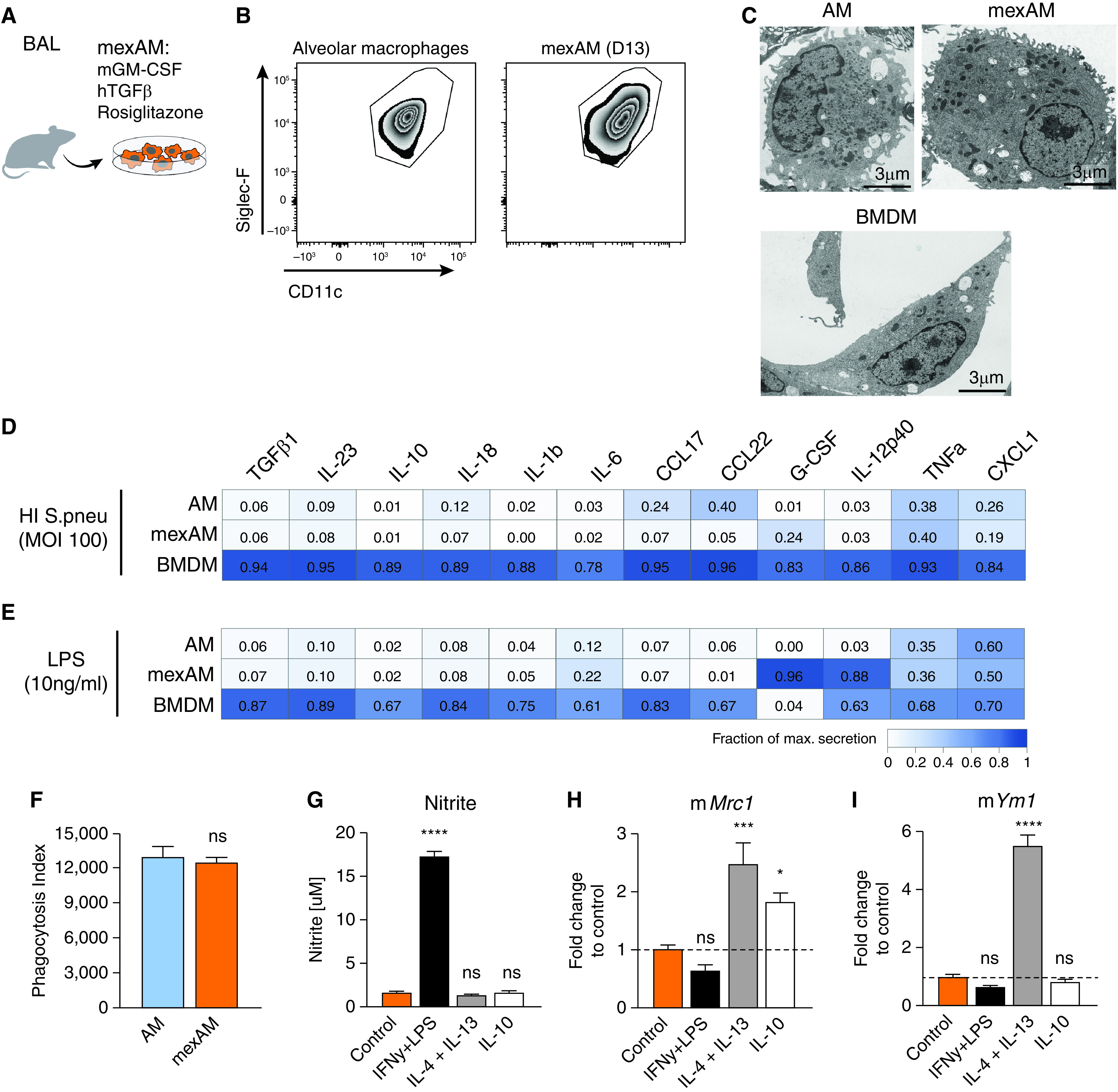
Figure 2.

MexAMs are functionally similar to primary AMs. (A) Experimental setup. (B) FACS analysis of Siglec-F and CD11c expression on primary AMs and mexAMs on Day 13. Pregated on viable CD45+ cells. (C) Electron microscopy pictures of primary AMs, mexAMs, or BMDMs. Scale bars, 3 µm. (D and E) Measurement of indicated cytokines upon heat-inactivated S. pneumoniae (MOI 100) (D) or LPS (10 ng/ml) (E) stimulation of AMs, mexAMs, or BMDMs for 16 hours, expressed as fraction of maximal secretion. (F) Phagocytosis index of primary AMs and mexAMs. (G) Nitrite concentration in supernatants of polarized mexAMs after 16 hours. (H and I) M2 polarization markers mMrc1 (H) and mYm1 (I) assessed by qPCR in polarized mexAMs after 1.5 hours. (D–I) Graphs show means ± SEM of 3–4 biological replicates (D and E) or technical quadruplicates (F–I). Data are representative of at least two independent experiments. *P < 0.05, ***P < 0.001, and ****P < 0.0001 (one-way ANOVA followed by Dunnett’s multiple comparison test). BMDMs = bone marrow-derived macrophages; HI = heat-inactivated; mexAM = mouse ex vivo cultured alveolar macrophages; MOI = multiplicity of infection; ns = not significant; S.pneu = Streptococcus pneumoniae.
