Figure 4.

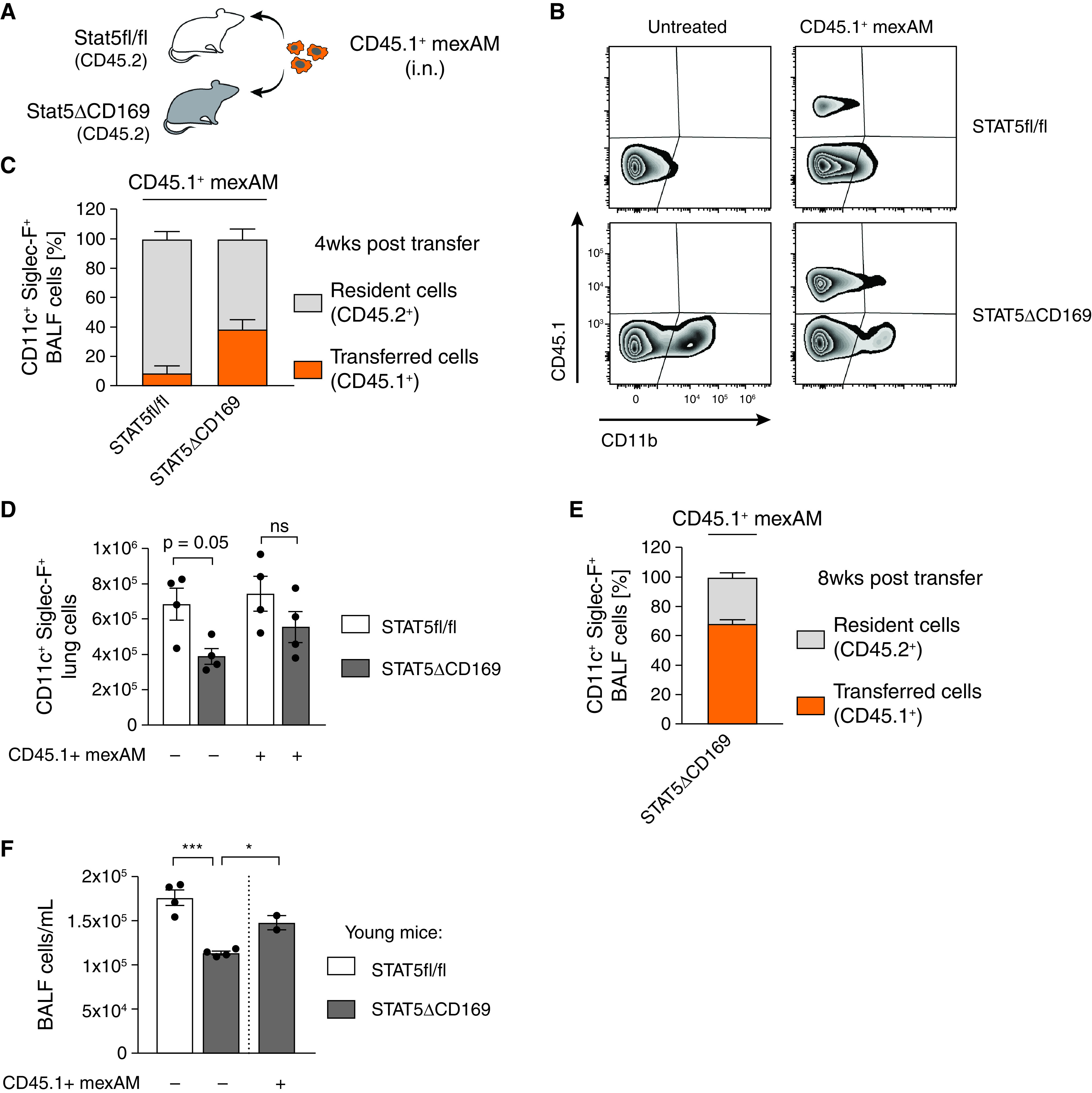
MexAMs engraft efficiently in a partially depleted AM niche in vivo. (A) Experimental setup. Intranasal transfer of CD45.1+ mexAMs into CD45.2-expressing control (STAT5fl/fl) or STAT5ΔCD169 mice. (B) FACS analysis of CD45.1 and CD11b expression of control (upper panel) and STAT5ΔCD169 (lower panel) BAL fluid (BALF) cells untreated, or 4 weeks after transfer of CD45.1+ mexAMs. Pregated on viable Siglec-F+/CD11c+ cells. (C) Percentage of resident (CD45.2+, gray) and transferred (CD45.1+, orange) cells in BALF of control (STAT5fl/fl, n = 4) and STAT5ΔCD169 mice (n = 4) 4 weeks after CD45.1+ mexAM transfer. (D) CD11c+Siglec-F+ lung cell number in STAT5fl/fl and STAT5ΔCD169 mice untreated and 4 weeks after transfer of CD45.1+ mexAMs. (E) Percentage of resident (CD45.2+, gray) and transferred (CD45.1+, orange) cells in BALF of STAT5ΔCD169 mice (n = 4–5) 8 weeks after CD45.1+ mexAM transfer. (F) BALF cell count per ml in STAT5fl/fl and STAT5ΔCD169 mice 14 weeks after transfer of CD45.1+ mexAMs into young (14 d) mice. Graphs show means ± SEM of 2–5 biological replicates. *P < 0.05, and ***P < 0.001 (one-way ANOVA followed by Sidak’s multiple comparison test). i.n. = intranasal.
