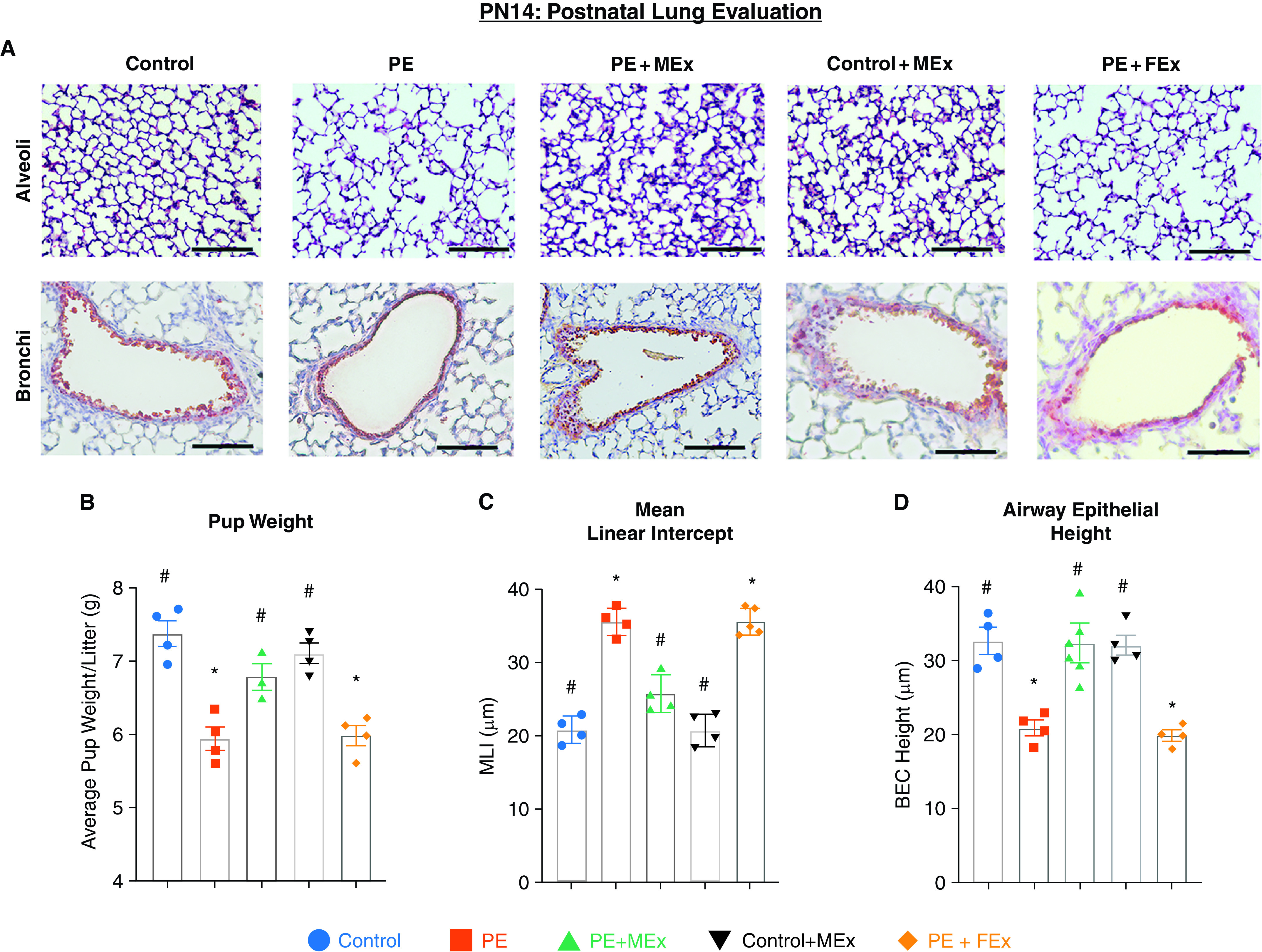
Figure 3.

Preeclampsia-associated disruptions in postnatal alveolar and bronchial epithelial morphology are ameliorated by antenatal MEx therapy. (A) Representative hematoxylin and eosin images of alveoli and bronchi from PN14 lung histology showing disrupted alveolarization and abnormal airway epithelium. Scale bars, 60 µm. (B) Pup weight analysis was determined at PN14. (C) Mean linear intercept analysis quantifying lung alveolarization. (D) Measurement of airway epithelial height was assayed by clara cell secretory protein (CCSP) staining in lung tissue sections. *#Statistically significant (P < 0.05) changes between experimental groups as determined by one-way ANOVA are denoted by different symbols. Data are representative of four independent experiments (n = 3–4 litters per experiment). BEC = bronchial epithelial cell; MLI = mean linear intercept.
