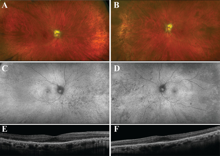
Figure 1. Multicolor wide-field photos, fundus autofluorescence (FAF), and optical coherence tomography (OCT) from patient with TULP1 cancer-associated retinopathy.
A, B: Multicolor wide-field photos showing fundus appearance of the right (A) and left (B) eye. Note attenuated retinal vessels and subtle pigmentary changes in the peripheral retina of both eyes.
C, D: FAF of the right (C) and left (D) eye. Note hypo-FAF speckling bilaterally in the peripheral retina.
E, F: Spectral domain OCT revealed loss of outer retinal layers outside the fovea in the right (E) and left (F) eye.

