. 2021 Dec 1;82(2):334–345. doi: 10.1158/0008-5472.CAN-21-2843

Figure 2.

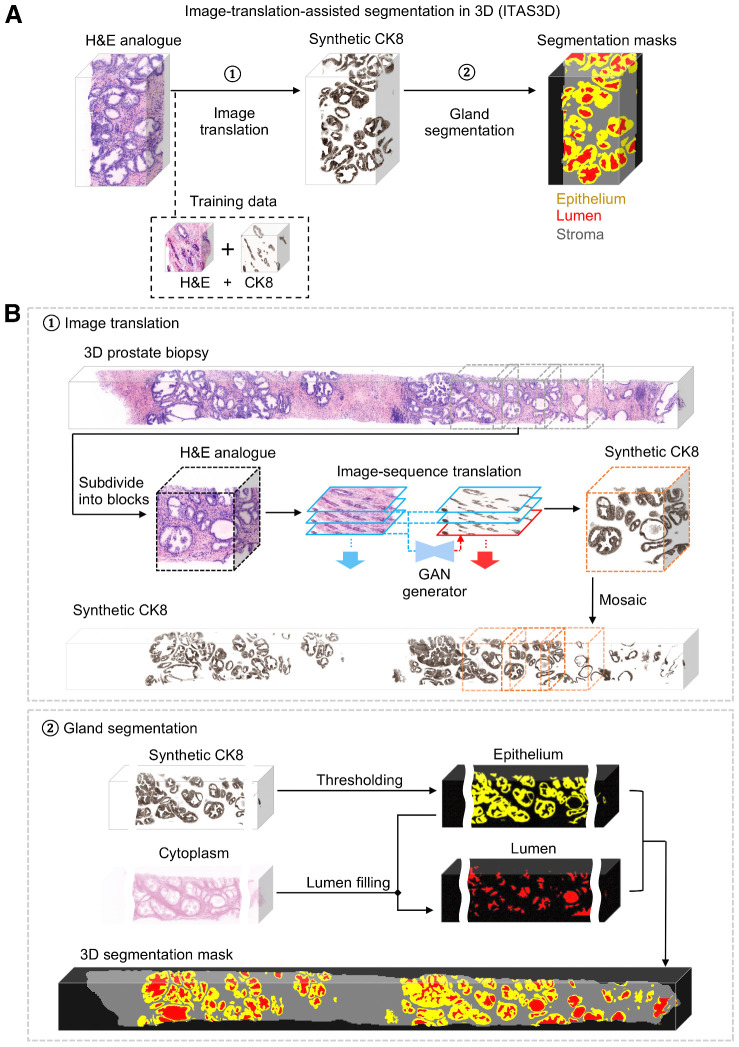
Figure 2. ITAS3D: a two-step pipeline for annotation-free 3D segmentation of prostate glands. A, In step 1, a 3D microscopy dataset of a specimen, stained with a rapid and inexpensive fluorescent analogue of H&E, is converted into a synthetic CK8 immunofluorescence dataset by using an image-sequence translation model that is trained with paired H&E analogue and real-CK8 immunofluorescence datasets (tri-labeled tissues). The CK8 biomarker, which is utilized in standard-of-care genitourinary pathology practice, is ubiquitously expressed by the luminal epithelial cells of all prostate glands. In step 2, traditional computer-vision algorithms are applied to the synthetic-CK8 datasets for semantic segmentation of the gland epithelium, lumen, and surrounding stromal regions. B, In step 1, a 3D prostate biopsy is subdivided into overlapping blocks that are each regarded as depth-wise sequences of 2D images. A GAN-trained generator performs image translation sequentially on each 2D level of an image block. The image translation at each level is based on the H&E analogue input at that level while leveraging the H&E analogue and CK8 images from two previous levels to enforce spatial continuity between levels (i.e., a “2.5D” translation method). The synthetic-CK8 image-block outputs are then mosaicked to generate a 3D CK8 dataset of the whole biopsy to assist with gland segmentation. In step 2, the epithelial cell layer (epithelium) is segmented from the synthetic-CK8 dataset with a thresholding-based algorithm. Gland lumen spaces are segmented by filling in the regions enclosed by the epithelia with refinements based on the cytoplasm channel (eosin fluorescence). See Supplementary Methods for details.

ITAS3D: a two-step pipeline for annotation-free 3D segmentation of prostate glands. A, In step 1, a 3D microscopy dataset of a specimen, stained with a rapid and inexpensive fluorescent analogue of H&E, is converted into a synthetic CK8 immunofluorescence dataset by using an image-sequence translation model that is trained with paired H&E analogue and real-CK8 immunofluorescence datasets (tri-labeled tissues). The CK8 biomarker, which is utilized in standard-of-care genitourinary pathology practice, is ubiquitously expressed by the luminal epithelial cells of all prostate glands. In step 2, traditional computer-vision algorithms are applied to the synthetic-CK8 datasets for semantic segmentation of the gland epithelium, lumen, and surrounding stromal regions. B, In step 1, a 3D prostate biopsy is subdivided into overlapping blocks that are each regarded as depth-wise sequences of 2D images. A GAN-trained generator performs image translation sequentially on each 2D level of an image block. The image translation at each level is based on the H&E analogue input at that level while leveraging the H&E analogue and CK8 images from two previous levels to enforce spatial continuity between levels (i.e., a “2.5D” translation method). The synthetic-CK8 image-block outputs are then mosaicked to generate a 3D CK8 dataset of the whole biopsy to assist with gland segmentation. In step 2, the epithelial cell layer (epithelium) is segmented from the synthetic-CK8 dataset with a thresholding-based algorithm. Gland lumen spaces are segmented by filling in the regions enclosed by the epithelia with refinements based on the cytoplasm channel (eosin fluorescence). See Supplementary Methods for details.