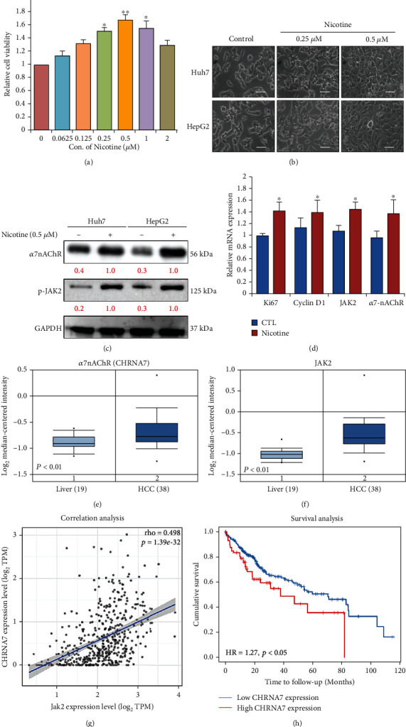
Figure 1.

Nicotine treatment promotes cell proliferation of HCC cells. (a) Huh7 and HepG2 cells (5 × 10⁴ cells) were treated with the indicated concentrations of nicotine for 48 h. The cell viability of Huh7 and HepG2 was determined by the CCK-8 assay. (b) Optical microscopy showed nicotine treatment at 0.25-0.5 μM dose-dependently induced morphological changes of HCC (Huh7 and HepG2) cells. Scale bar 100 μm. (c) Huh7 and HepG2 cells stimulated with 0.5 μM nicotine for 48 h were harvested, and the effects of nicotine on α7nAChR and JAK2 expression were determined by western blot analysis. (d) Real-time PCR analysis of Ki67, Cyclin D1, JAK2, and α7nAChR mRNA in HCC cells. (e, f) Expression of α7nAChR (CHRNA7) and JAK2 in liver cancer (GSE14323). (g) Correlation analysis of α7nAChR (CHRNA7) expression with the JAK2 expression in LIHC TCGA patient data. (h) Kaplan-Meier analysis of expression of α7nAChR (CHRNA7) and JAK2 for LIHC patients based on the result from the public TCGA data. GAPDH was used as an internal control. Bar represent the mean ± SD values of three independent experiments; ∗p < 0.05; ∗∗p < 0.01.
