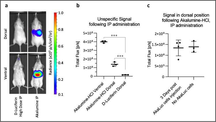
Fig. 6.
In the absence of cells, Akalumine-HCl generates a non-specific signal from the liver when administered IP. High-dose D-Luciferin or Akalumine-HCl was injected IP in vivo. The mice were then imaged in dorsal (23 min post substrate administration) and ventral (20 min post administration) positions, with no emission filter, a 22.8 field of view, a f-stop of 1, a binning of 8 and an exposure time of 180 s. a Representative images of the mice following substrate administration (radiance scale from 1 × 10⁴ to 1 × 10⁵ p/s/cm2/sr). b Quantification of the non-specific signal detected in the liver region of the mice injected with Akalumine-HCl IP, analysed both in ventral and dorsal position, compared with the signal coming from the liver region of mice injected with high dose D-Luciferin IP. Data are displayed as mean ± SD from n = 3. Statistical analysis performed using a one-way ANOVA and Tukey’s multiple comparison post hoc test. ***p < 0.001 post substrate administration. c Comparison of the signal detected in the liver of mice injected with Akalumine-HCl, three days post administration of 2.5 × 10⁵ AkaLuc expressing cells IV (n = 4) vs. the non-specific luminescence from naive mice that received Akalumine-HCl alone (n = 3), 20 min. There is no statistically significant difference between the two groups (unpaired t-test). Data are displayed as mean ± SD

