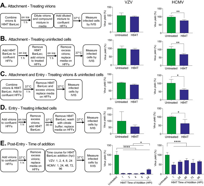
Figure 2.
Effects of H84T BanLec on extracellular VZV and HCMV virions and on infected cells. Each experimental condition tests a different aspect of H84T BanLec’s mechanism of action including attachment (A, B), attachment and entry (C), entry (D), and post-entry steps (E). Cell-free virions and H84T BanLec were added to HFF cell monolayers based on the experimental design for each condition. Cell-free VZV was prepared from a fresh culture of infected HFF cells; the MOI was approximately 0.01. HCMV was prepared from a frozen, titered stock; the MOI was 0.05. H84T BanLec was used at a concentration of 100 nM for VZV and 2 µM for HCMV. (A) Virions were mixed with H84T BanLec on ice for 1 h, and were then diluted 1:50 for VZV and 1:1000 for HCMV prior to adding to HFFs. (A–D) Infected cells were measured by bioluminescence imaging 24 h post-infection for VZV, and 7 days post-infection for HCMV. (E) Infected cells were measured by bioluminescence imaging 48 h post-infection for VZV, and 7 days post-infection for HCMV. Virus yield (%) was calculated from the average Total Flux of untreated wells divided by the Total Flux of each well. Bars and error bars represent the mean + SD. N = 4 biological replicates. Asterisks indicate significance [*p < 0.05, **p < 0.01, ****p < 0.0001, p < 0.05, Student’s t-test (A–D) or one-way ANOVA with Dunnett’s post hoc test (E)].

