Figure 3.
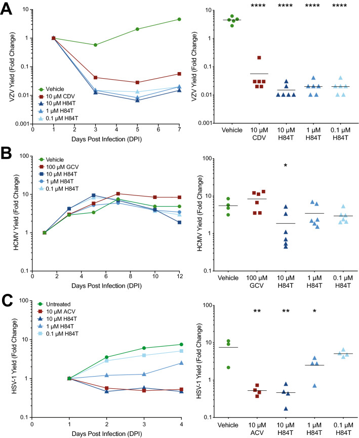
H84T BanLec evaluation in skin organ culture. Adult skin explants were inoculated with VZV (A), HCMV (B), or HSV-1 (C) and then placed on NetWells over medium that contained H84T BanLec or positive control antiviral compounds (cidofovir, CDV, 10 µM; ganciclovir, GCV, 100 µM; acyclovir, ACV, 10 µM). Virus spread was measured by bioluminescence imaging starting on 1 DPI. Virus yield on subsequent days was calculated by dividing the average Total Flux (photons/sec/cm2/steradian) by the average Total Flux on 1 DPI. Viral growth kinetics (left panels, each symbol is the average for the group) were analyzed for statistical significance on the last day (right panels), where each symbol represents one piece of skin and the bar is the mean of the group. Asterisks indicate significance between the treated groups and vehicle, [*p < 0.05, **p < 0.01, ****p < 0.0001, p < 0.05, one-way ANOVA, Dunnett’s (A, C) or Dunn’s (B) post hoc test]. N = 4–6 biological replicates.

