Figure 2.
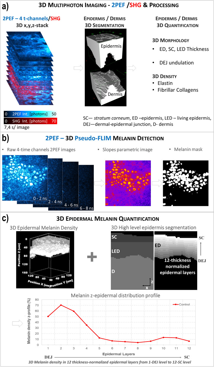
Global analysis process for in vivo 3D multiphoton images allowing 3D skin automatic layers segmentation and constituents quantification. (a) The first step of global 3D analysis of z-stacks of combined 2PEF-FLIM (4 time channels)/SHG images briefly consists in identifying the epidermal and dermal layers (3D automatic segmentation) and quantifying their morphology (thickness, DEJ 3D-shape) and the 3D dermal density of elastin and fibrillar collagens. (b) The z-stack of 2PEF-FLIM (4 time channels) images is further processed for melanin detection using Pseudo-FLIM method. The first 3-time channels are used for slope parametric image calculation and a melanin mask is obtained by applying a threshold to keep the high slopes values (above 70). (c) The 3D z-stack of melanin masks and the 3D automatic segmentation of the epidermis and its sub-layers are jointly used to estimate a 3D epidermal, SC or LED melanin density. By defining 12-thickness normalized epidermal layers, the epidermal melanin density z-distribution (z-profile of melanin density in the 12 thickness-normalized epidermal layers from 1—DEJ level to 12—SC level) can be assessed.

