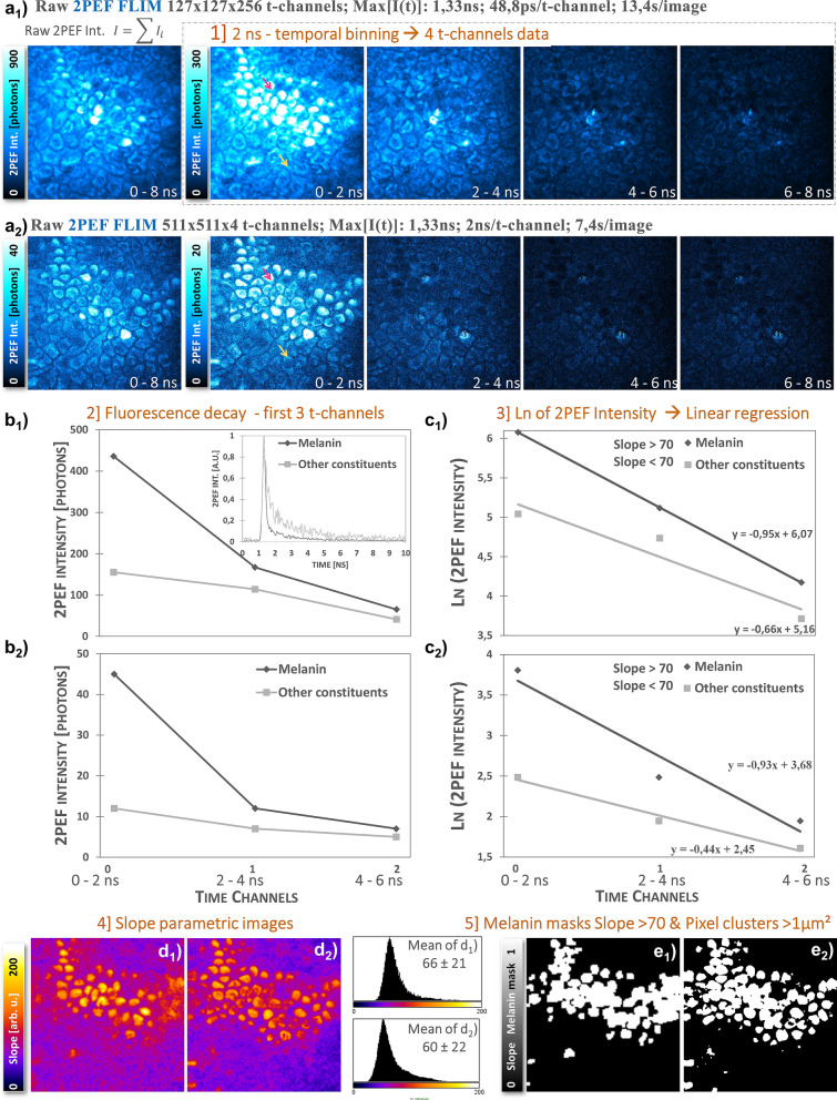
Figure 5.
Effect of image acquisition time on in vivo human skin Pseudo-FLIM melanin detection. (top) 2PEF intensity images within 0–8 ns time range and within the first 4 temporally binned time channels with 2 ns integration time. (a1) 2D 2PEF-FLIM image of 127 × 127 pixels × 256 time channels acquired within the basal layer of the epidermis (104 µs pixel dwell time); (a2) 2D 2PEF-FLIM image of 511 × 511 pixels × 4 time channels extracted from a z-stack and acquired almost within the same 2D plane at 28 µs pixel dwell time. (b1, b2) shows the corresponding 2PEF intensity decays of the first 3 temporally binned channels for a high slope (red arrow, melanin) and small slope (yellow arrow, other constituents) pixels. The full decay of these two pixels extracted from the 256 t-channels images is given in the insert in graph (b1). Graphs (c1, c2) show the natural logarithm transformation and linear regression fitting to extract the slopes of the decays. Images (d1, d2) show the corresponding slope parametric images and their Pseudo-FLIM melanin mask is given in images (e1, e2).

