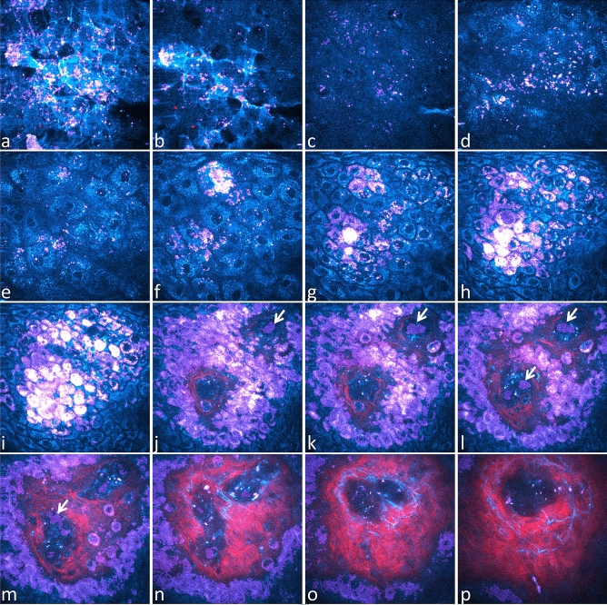
Figure 6.
In vivo 3D multiphoton images of human skin – acquisition of combined 2PEF-FLIM (4 time channels)/SHG z-stacks compatible with Pseudo-FLIM melanin detection. 2PEF intensity is shown in cyan hot color, SHG in red and Pseudo-FLIM melanin mask pixels in purple. High 2PEF signal intensities appear in white color. Images are extracted from a z-stack of 70 en face images acquired with 2.346 µm z-step. (a) stratum corneum disjunctum; (b) stratum corneum disjunctum/compactum interface; (c, d) compactum / granulosum interface; (e, f) stratum granulosum; (g, h) stratum spinosum; (i) stratum basale; (j–o) stratum basale/dermis interface; (p) superficial dermis. Within the blood capillaries, Pseudo-FLIM detects cells with high slope values, fast decays, that most probably emit 2PEF signals from hemoglobin (see arrows). As the image acquisition time is slower than the blood flow, some blood cells appear with a deformed shape as indicated by the arrow in image m.

