Figure 2.
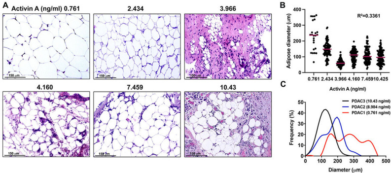
Serum activin A levels can be correlated with loss of visceral adipose tissue in PDAC patients. (A) H&E images of visceral adipose tissue from PDAC patients with corresponding activin A levels in ng/ml. Scale bar = 150 µm. (B) Measurement of adipocyte diameter in visceral adipose tissue from PDAC patients. The diameters of adipocytes were measured from several histological sections. 6 sections, 0.761 ng/ml; 14 sections, 2.434 ng/ml; 14 sections, 3.966 ng/ml; 11 sections, 4.160 ng/ml; 10 sections, 7.459 ng/ml; 6 sections, 10.425 ng/ml. Corresponding serum activin A levels are included in ng/ml. (C) Frequency curve of adipocyte diameter in visceral adipose tissue from patients with low serum activin A (0.761 ng/ml, red line), elevated serum activin A (8.984 ng/ml, blue line), and increasingly elevated serum activin A (10.425 ng/ml, black line). The diameters of adipocytes were measured with 6 histological sections each.

