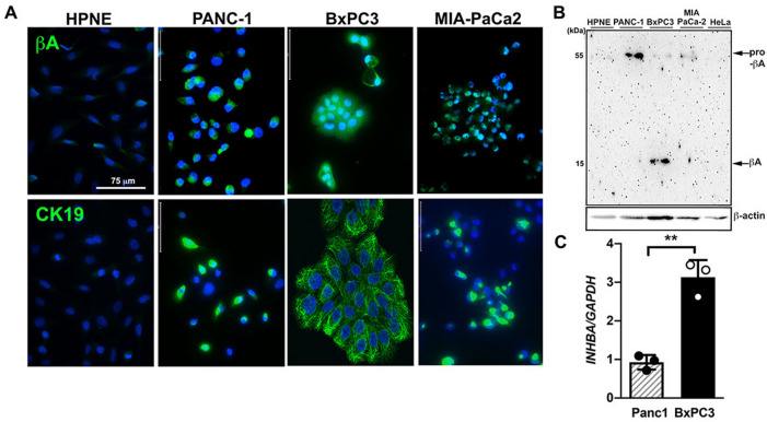
Figure 3.
Activin A is highly expressed in tumor-derived cell lines from PDAC patients. (A) Immunofluorescence images of activin A (βA, top panels, AF488) and cytokeratin 19 (CK19, bottom panels, AF488) in hTERT-HPNE, PANC-1, BxPC-3 and MIA PaCa-2 cells. Nuclei were stained with DAPI. Scale bar = 75 µm. (B) Immunoblotting analysis of activin A expression in hTERT-HPNE, PANC-1, BxPC3, MIA PaCa-2 and HeLa cell lysates. (C) Relative INHBA mRNA expression levels in PANC-1 and BxPC-3 cells.

