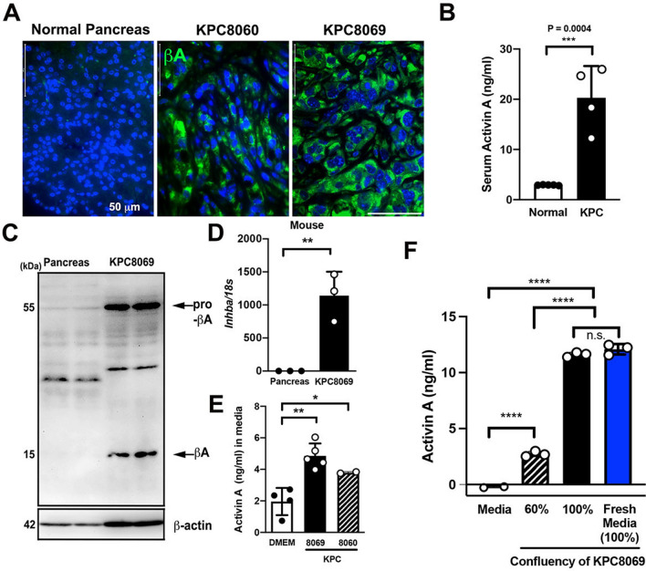
Figure 4.
Activin A is highly expressed in tumor specimens and tumor-derived cell lines of genetically engineered mouse models of PDAC. (A) Immunofluorescence images of activin A (AF488) in normal mouse pancreas tissue and tumor specimens of KPC 8060 and KPC 8069 mice. Scale bar = 50 µm. (B) Measurement of serum activin A levels in healthy control C567BL/6 J and KPC mice. (C) Immunoblotting analysis of activin A expression in healthy control C57BL/6 J mouse pancreas tissue and KPC8069 cell lysates. (D) Relative Inhba mRNA expression levels in healthy control C57BL/6 J mouse pancreas tissue and KPC8069 cells. (E) Measurement of activin A concentration in CM from KPC8060 and KCP8069 cells along with RPMI-1640. (F) Measurement of activin A concentration in CM from KPC8069 cells at 60% confluency, 100% confluency and 100% confluency following reintroduction (a blue bar) of fresh culture medium.

