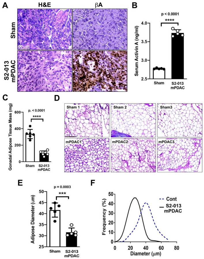
Figure 5.
Activin A can be correlated with loss of gonadal visceral adipose tissue in an orthotopic implantation model of PDAC. (A) Representative immunohistochemical images of activin A expression in healthy control C57BL/6 J pancreatic tissue and tumor specimens of S2-013 mPDAC. Scale bar = 50 µm. (B) Measurement of serum activin A levels in sham and S2-013 mPDAC. (C) Mass of gonadal fat pad. (D) H&E images of adipose tissue from the gonadal fat pad in sham and S2-013 mPDAC. Scale bar = 150 µm. (E) Measurement of adipocyte diameter in gonadal adipose tissue from sham and S2-013 mPDAC. The diameters of adipocytes were measured from several histological sections (n = 5). The average adipocyte diameter from each animal was marked with a dot. F. Frequency curve of adipocyte diameter in gonadal fat pad of sham mice (dashed line) and S2-013 mPDAC (solid line).

