Figure 6.
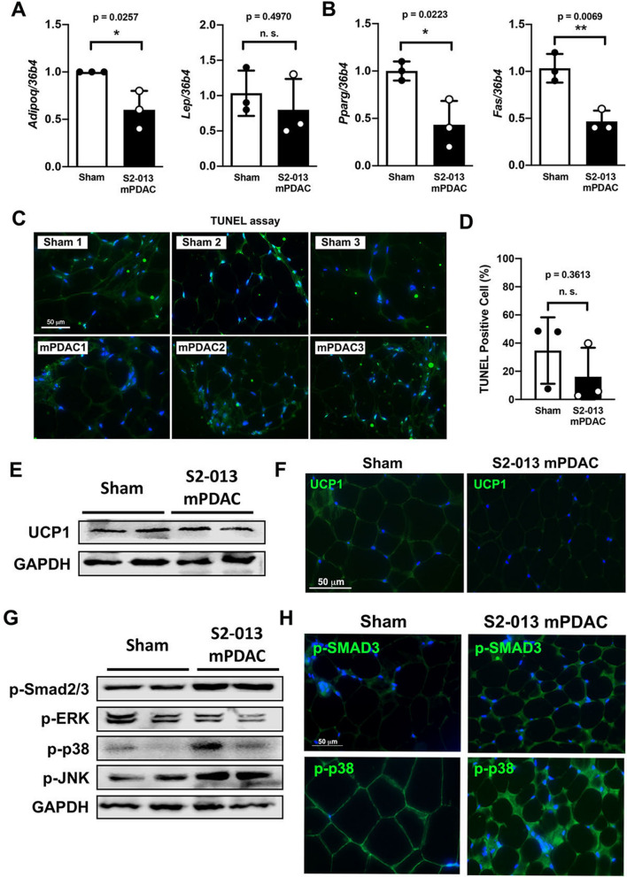
Visceral adipose tissue remodeling in an orthotopic implantation model of PDAC does not rely on increased browning of white adipose tissue. (A) Relative mRNA expression levels of Adipoq and Lep in gonadal adipose tissue from sham and S2-013 mPDAC. (B) Relative mRNA expression levels of Pparg and Fas in gonadal adipose tissue from sham and S2-013 mPDAC. (C) TUNEL assay with adipose tissue from the gonadal fat pad of sham (n = 3) and S2-013 mPDAC (n = 3). (D) Area percentage of green pixels indicating TUNEL-positive signals as measured by Image J. Each dot represents the percentage of TUNEL-positive signals in the gonadal fat pad of one animal. (E) Representative immunofluorescence image of UCP-1 expression in the gonadal fat pad of sham control and S2-013 mPDAC. Nuclei were stained with DAPI. (F) Immunoblotting with UCP1 in gonadal adipose tissue from sham and S2-013 mPDAC. (G) Immunoblotting with the markers for canonical activin A (p-Smad2/3) and ERK/MAPK pathways in adipose tissue from the gonadal fat pad of sham and S2-013 mPDAC. (H) Representative immunofluorescence images of p-SMAD3 and p-p38 expression in adipose tissue from the gonadal fat pad of sham and S2-013 mPDAC. Nuclei were stained with DAPI. Scale bar = 50 µm for all microscope images.

