Figure 1.

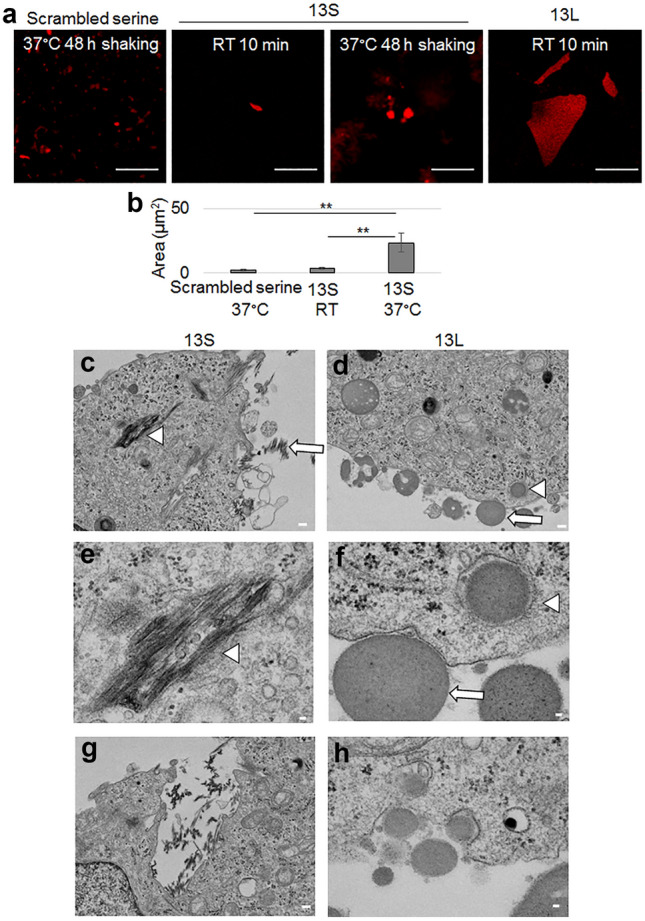
Aggregated 13S and 13L enter PC12 cells. (a,b) Fluorescent images of clusters of aggregates of TAMRA-labeled 13S and 13L peptides (1 mg/ml in water, red). 13S and 13L were incubated for 10 min at room temperature. 13S and scrambled serine were also incubated for 48 h at 37 °C with shaking (a). Area of aggregates were quantified (n = 20 each) (b). ANOVA, **p < 0.01. Scale bar, 20 µm. (c–h) Transmission electron microscopic images of undifferentiated PC12 cells having 13S or 13L. 13S peptide (1 mg/ml in water) was incubated for 48 h at 37 °C with shaking. Then, the aggregated 13S peptide was added in the culture medium of PC12 cells to be at a final concentration of 10 µg/ml. The stock solution of 13L peptide was directly applied in the culture medium to be at a final concentration of 10 µg/ml. The cells having two peptides were incubated for 1 day at 37 °C. Extracellular aggregates (arrows) of 13S (c) and 13L (d) are shown. 13S (c,e,g) and 13L (d,f,h) aggregates inside the cells (arrowheads) are also shown. The invaginated vesicles having aggregates inside are also shown (g, 13S; h, 13L). Scale bars, 20 µm (a), 1 µm (c,d,g), 200 nm (e,f,h).
