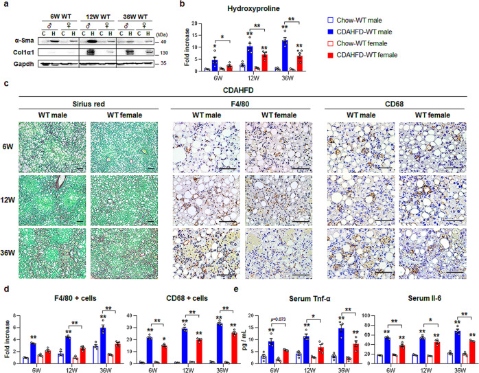
Fig. 2. Enhanced liver fibrosis and inflammation in CDAHFD-fed male than female.
a Western blot analysis of hepatic alpha-smooth muscle actin (α-Sma) and collagen 1 alpha 1 (Col1α1) in wild-type (WT) male and female mice treated with chow or choline-deficient, L-amino acid-defined, high-fat diet (CDAHFD). Each lane contains protein lysates pooled from representative three mice per group with equal concentration. Glyceraldehyde 3-phosphate dehydrogenase (Gapdh) was used as internal control. The data shown represent one of three experiments with similar results. b Hepatic hydroxyproline content in liver tissues from representative mice per each group. Data represent the mean ± S.E.M. (n ≥ 4/group, *p < 0.05, **p < 0.005 vs own control). c Representative images of Sirius red- (left panel) and F4/80- (middle panel) and CD68-stained (right panel) liver sections from the CDAHFD-WT groups (Scale bar, 50 μm). d Quantitative F4/80- or CD68-stained data from these WT mice. F4/80- or CD68-positive Kupffer cells were quantified by dividing the total numbers of positive cells by the total number of Kupffer cells. Data represent the mean ± S.E.M. (n ≥ 4/group, *p < 0.05, **p < 0.005 vs own control). e Levels of serum Tnf-α and Il-6 in representative mice from each group. Data represent the mean ± S.E.M. (n ≥ 4/group, *p < 0.05, **p < 0.005 vs own control). Gray circles represent individual data points. See Supplementary Data for statistical details.

