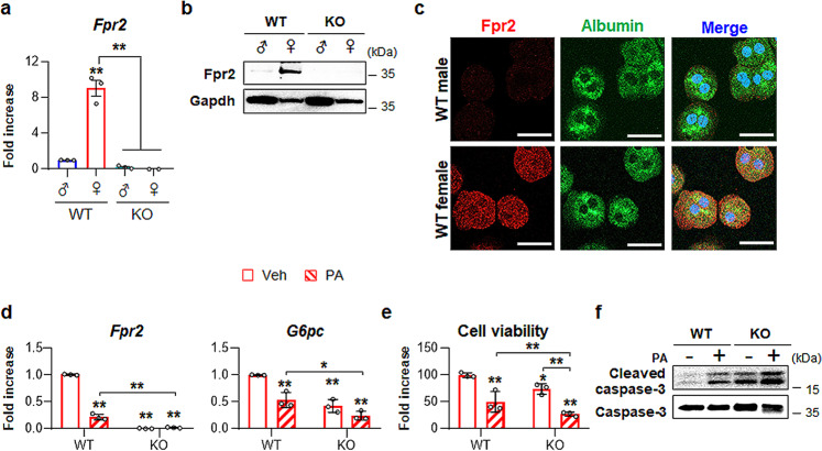
Fig. 7. Higher expression of Fpr2 in the livers of female mice is related with hepatocyte protection.
a qRT-PCP analysis for Fpr2 expression in primary hepatocytes (pHEPs) from WT (male n = 2, female n = 2) and KO mice (male n = 2, female n = 2). Total eight mice were employed in each hepatocyte isolation, and the experiments were replicated at least three times and the mean ± S.E.M. results are graphed (*p < 0.05, **p < 0.005 vs WT male-pHEPs). b Western blot analysis and c double immunofluorescent images of Fpr2 (red) with albumin (green) in these cells. Gapdh was used as internal control. DAPI (blue) was used as nuclear counterstaining. Data shown represent one of three experiments with similar results (Scale bar, 20 μm). d qRT-PCR analysis for Fpr2 and glucose-6-phosphatase (G6pc) in, e cell viability of, and f western blot analysis of cleaved Caspase-3 and pro Caspase-3 in WT and KO female mice-isolated pHEPs treated with vehicle (Veh) or 250 μM of palmitate (PA). The data shown represent one of three experiments with similar results. The mean ± S.E.M. results obtained from three repetitive experiments are graphed (*p < 0.05, **p < 0.005 vs WT-Veh). Gray circles represent individual data points. See Supplementary Data for statistical details.

