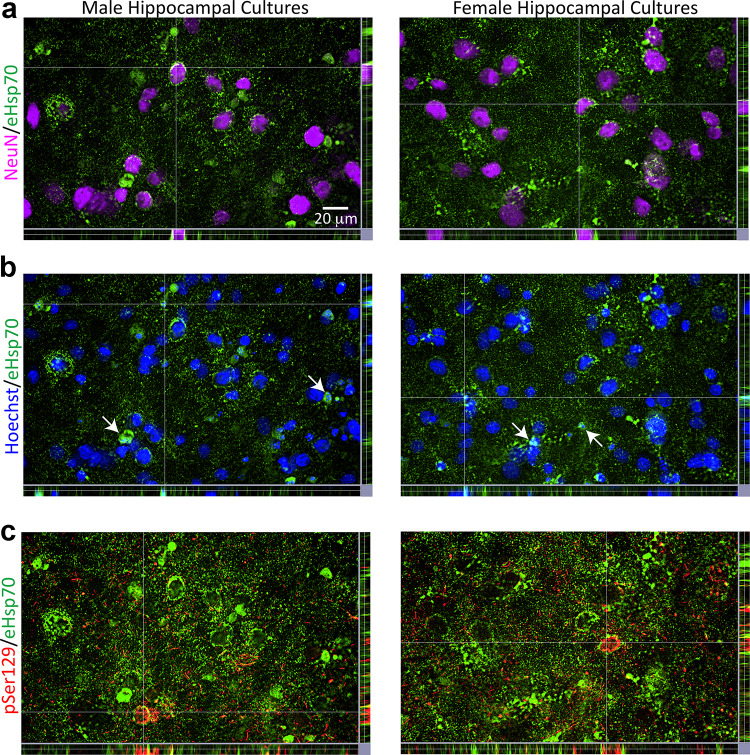
Fig. 4.
Uptake of eHsp70 by male and female primary hippocampal cells. Primary hippocampal cultures harvested from male vs. female rat pups were exposed to 1 μg/mL α-synuclein fibrils and 75 µg/mL of eHsp70 for 10 days or their respective vehicles (not shown). Representative z-stack views in a show eHsp70 encompassing NeuN+ neuronal nuclei in both male and female cultures. Representative z-stack views in b show the eHsp70 within or surrounding shrunken Hoechst+ nuclei. Arrows in b point to additional examples outside of the point of intersection. In the z-stack images of panel c, eHsp70 immunolabel was brightened to show parts of perinuclear pSer129+ inclusions caged in by eHsp70. Photomontages in a–c are all captured from the same field of view

