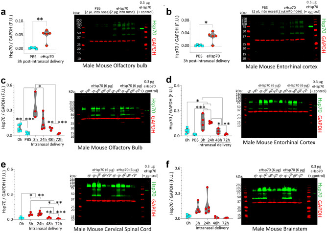
Fig. 6.
eHsp70 enters the male mouse brain from the nasal cavities within 3 h and dissipates by 72 h post-infusion. a–b Three-month-old male mice were anesthetized with isoflurane and infused with low-endotoxin eHsp70 (2 µg; Enzo) or an equivalent volume of PBS (2 µL) behind the nares, after which the mice were left supine for 10 min and then returned to home cages. Three hours post-infusion, brains were extracted, and the olfactory bulb (a) and entorhinal cortices (b) were subjected to immunoblotting with antibodies against human Hsp70. As a positive control, recombinant human eHsp70 protein was loaded directly into the second-to-last lane of the gel. MW standards were included on both sides of all gels (red ladders). Only the band at ~ 70 kDa was quantified in the violin plots. Higher bands may be aggregated eHsp70 or nonspecific, and the lower bands of ~ 44 kDa are likely to be the cleaved ATPase domain. For studies determining eHsp70 uptake and clearance in older (17–20 months old) male mice, 6 µg of low-endotoxin eHsp70 (Enzo) was intranasally infused and the olfactory bulb (c), entorhinal cortex (d), cervical spinal cord (e), and brainstem (f) were subjected to immunoblotting. For immunoblots in c–f, the positive control (recombinant eHsp70 protein) was loaded in the last lane of the gel. Brains were collected at 0 h (sham control), 3 h, 24 h, 48 h, and 72 h post-infusion. The younger PBS controls from a–b were also included in the blots shown in c–d. For the 3 h group in c, we began with n = 3 male mice but an additional n was added by the time the data in d–f were collected. The spinal cord from one male mouse in the 0 h group in e was lost during dissections. * p ≤ 0.05, ** p ≤ 0.01, *** p ≤ 0.001. Data in panel a were analyzed by the two-tailed Mann–Whitney U. Data in panel b were analyzed by the two-tailed unpaired t-test with Welch’s correction. Data in panels c–f were analyzed by the one-way ANOVA followed by Bonferroni post hoc

