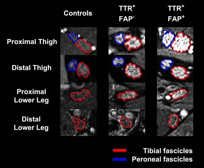
Fig. 2.
Nerve lesion detection and anatomical lesion distribution in ATTRv amyloidosis. The tibial (encircled in red) and peroneal fascicles (encircled in blue) within the sciatic nerve and the distal continuation as tibial nerve are shown on representative axial fat-saturated T2w images. A marked increase in multifascicular T2w hyperintense nerve lesions was observed in asymptomatic varTTR-carriers (TTR+ FAP−) compared to healthy controls whose nerves appear with an intermediate T2w signal similar to the surrounding muscles. In symptomatic ATTRv amyloidosis (TTR+ FAP+) nerve lesions showed an additional significant increase in T2w hyperintensity compared to the varTTR-carriers. A marked increase in nerve caliber measured as CSA was also already observable in the asymptomatic group and was even more pronounced in symptomatic ATTRv amyloidosis. The spatial lesion distribution appears to be diffuse, affecting the whole nerve cross-section. However, a clear proximal-to-distal gradient of nerve lesions was found in both groups with predominant lesions within the sciatic nerve at thigh level, indicating that nerve injury in distally-symmetric PNP in ATTRv amyloidosis starts and progresses in proximal sections of the PNS. From Kollmer et al. [53] under the Creative Commons Attribution license

