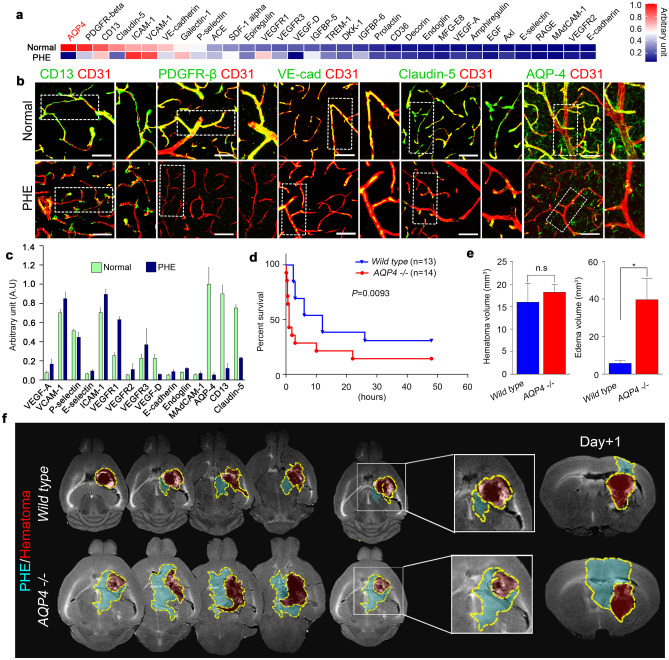
Fig. 2.
Decreased AQP4 expression in PHE is associated with poor survival outcomes. a Protein array of tissues from normal and PHE sites. b Representative confocal microscopic images of vascular-associated molecules in normal and PHE tissues (magnification × 20). c Quantification of vascular-associated proteins in the PHE region. d Survival curves of wild-type and AQP4−/− mice after intracerebral hemorrhage. e Hematoma volumes and edema volumes of wild-type and AQP4−/− mice after intracerebral hemorrhage. f MR images showing the chronological changes of PHE and hematoma in wild-type and AQP4−/− mice. White scale bar, 50 μm. Data are mean ± SD. *P < 0.05. *Data in a, b, c, and e were obtained at post-ICH day 3

