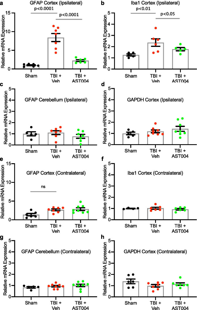
Fig. 3.
TBI-induced increases in mRNA levels of GFAP and Iba1 in the cortex are reduced by AST-004 treatments. Mice underwent sham or TBI and received AST-004 treatment (0.22 mg/kg) or vehicle 30 min post-injury. qRT-PCRs of brain homogenates were performed 7 days post-injury. mRNA levels for GFAP and Iba1 in the ipsilateral cortex are shown in histogram plots, as labeled a, b. GFAP mRNA levels in the ipsilateral cerebellum were not affected by TBI c GAPDH mRNA levels in the ipsilateral cortex were also unaffected d. For comparison, contralateral mRNA levels for GFAP, Iba1, and GAPDH in the cortex and GFAP in the cerebellum are also not significantly different in TBI-induced brain samples e, h. Number of mice used for sham: 3 males and 3 females, for TBI + vehicle: 3 males and 5 females, for TBI + AST-004: 5 males and 3 females. All samples run in duplicate. Brain samples from the contralateral side of the brain to the site of injury did not exhibit changes in mRNA levels for GFAP (cortex and cerebellum), Iba1 or GAPDH either after TBI only or TBI + AST-004 treatment AQ8(Fig. 3e–h)

