FIGURE 2.
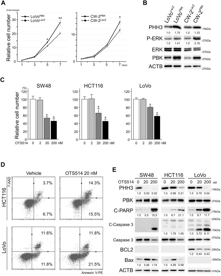
PBK enhanced the cellular proliferation of CRC cells. (A), exogenous PBK expression enhanced the cellular proliferation of CRC cells. Data are shown as mean ± S.D. *, p < 0.05, **, p < 0.01. (B), immunoblot analysis showing up-regulated P-ERK and PHH3 in PBK-induced CRC cells. (C), OTS514, a selective PBK inhibitor, suppressed the cellular proliferation of CRC cells. Data are shown as mean ± S.D. *, p < 0.05. (D), Annexin V assay in HCT116 and LoVo cell lines showing induction of apoptosis after OTS514 treatment. (E), protein expression of PHH3 and apoptosis-related proteins in CRC cells after OTS514 treatment. Note that BCL2 was expressed at under detectable levels in SW48 and HCT116. Bax expression was under detectable level in LoVo. Numbers below the immunoblot bands indicate relative expression.

