FIGURE 4.
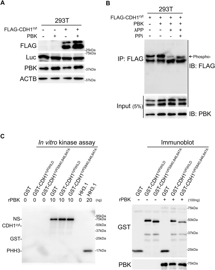
PBK accumulated CDH1 of phosphorylated form and phosphorylated histone H3. A and B, immunoblot analysis showing PBK accumulated CDH1cyt with phosphorylation. Note that the expression levels of co-transfected Luc were not affected by PBK. (C), in vitro kinase assay showing that both GST-tagged CDH1cyt/WILD and CDH1cyt/S840,846,847A were not phosphorylated by recombinant PBK (rPBK). In contrast, rHH3.1 was directly phosphorylated by rPBK (left panel). Immunoblot analysis showing CDH1cyt/WILD and CDH1cyt/S840,846,847A with or without kinase reaction. Note that no band shift was detected after kinase reaction (right panel). NS, non-specific.

