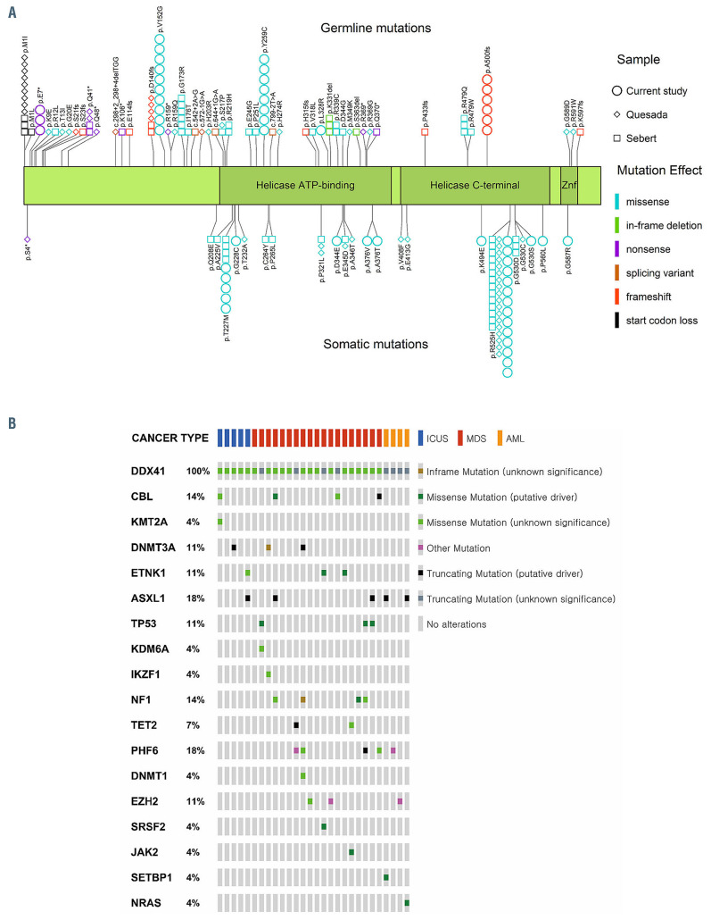
Figure 2.
Distribution of DDX41 mutations and concurrent mutations in other genes. (A) Distribution of DDX41 mutations detected in the current study and two previous studies (Quesada et al.10 and Sebert et al.11). This figure shows the differences in positional distribution (N-terminal skewed vs. C-terminal skewed) and mutational effects (variable vs. missense-dominated) between germline and somatic mutations. The protein structure of DDX41 was based on the RefSeq accession number of NM_016222.3 and the UniProtKB entry of Q9UJV9: the 622 amino acid long protein comprises the helicase ATP-binding domain (position 212-396), the helicase C-terminal domain (position 407-567), and a zinc finger domain (position 580-597). Different colors indicate different effects of mutations: light blue, missense mutation; light green, inframe indel; purple, nonsense mutation; brown, splicing mutation; red, frameshift mutation; black, start codon loss. Different shapes represent the three studies: square, Sebert et al.11 diamond, Quesada et al.10 circle, current study. (B) Concurrent mutations of other genes identified in bone marrow samples from DDX41-mutated patients. The types of genetic alterations and diseases are presented in the legend.

