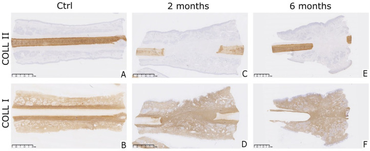
Figure 4.
Expression of collagen type II and I in tissue formed on the defect site. (A and B) Native nasal cartilage was positively stained for collagen type II and negatively for collagen type I in healthy native cartilage control samples. (C, D, E, and F) Collagen type I was abundantly present in the perichondrium, the mucosal and submucosal layers of both control and healing samples. Tissue formed on the place of the perforation was intensively stained brown indicating presence of collagen type I, while no positive staining for collagen type II was visible. Brown stain indicates positive staining. Scale bar represents 2.5 mm.

