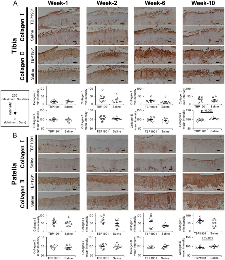
Figure 4.
TBP1901 intra-articular injections increased type II collagen expression at 10 weeks. (A) Immunohistochemical images with type I and II collagen and quantitative analyses in the tibia of the tibiofemoral (TF) joint. TBP1901 intra-articular injections significantly increased the type II collagen expression at week 10. (B) Immunohistochemical images with type I and II collagen and quantitative analyses in the patella of the patellofemoral joint. TBP1901 intra-articular injections significantly increased the type II collagen expression at week-10. The expressions of type I and type II collagen were analyzed by measuring the minimum and mean intensity values using ImageJ, respectively, on a scale from 0 (dark) to 255 (no staining). Values are the means in the TBP1901 and saline control groups at several time points (n = 8 each). P values were calculated using Welch’s t test. Magnification: ×100. Scale bar: 100 μm.

