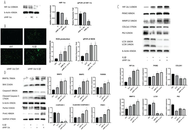
Figure 3.
Silencing HIF-1α inhibits cellular autophagic functions. C28/I2 chondrocyte cells were transfected with siRNA, and the subsequent (A) HIF-1α expression was evaluated using Western Blotting and qPCR of the treated cells. (B) The resulting cell ROS production due to silencing HIF-1α and IL1β insult was measured and quantified. (C) HIF-2α expression, glycolytic rate-limiting enzyme PKM2 expression, cell autophagic markers P62/LC3II and OA chondrocyte degeneration markers COL2A1/MMP13 were evaluated using Western blotting after silencing HIF-1α expression, and IL1β insult. (D) Mitophagic markers BNIP3/BAX and PINK/Parkin and apoptotic markers Caspase 3/Cleaved Caspase 3 were evaluated using Western Blotting in cells after HIF-1α silencing and IL1β insult. Average values were calculated from 3 individual experiments.

