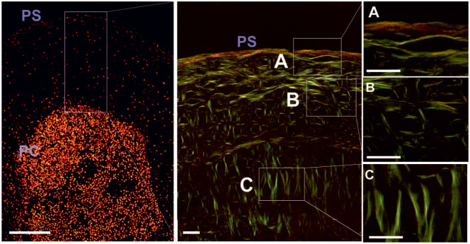
Figure 3.
Representative images of cell distribution and fiber orientation in the collagen matrix in chondroprogenitor (CP) pellets. Birefringent collagen fibers show developing zonation in the newly formed hyaline matrix and parallel to the pellet surface (PS) horizontal arranged birefringent fibers in the margin (A). Randomly organized collagen fibers were found in the central area (B). Perpendicularly oriented birefringent fibers (C) were located in proximity to the cell condensed center. Scale bar equals 30 µm, 400× magnification.

