Figure 3.
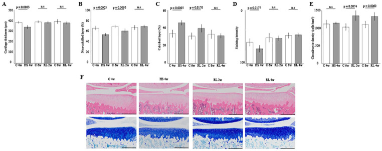
Histomorphometrical effect of the reloading on cartilage atrophy in the medial tibia: (A) cartilage thickness, (B) and (C) the percentage of the noncalcified and calcified layers in the articular cartilage, (D) matrix intensity of staining by toluidine blue, (E) chondrocyte density, and (F) representative histological findings. Scale bar = 500 µm. The thinning of the articular cartilage and decreases of staining intensity caused by disuse cartilage atrophy was restored by reloading for 2 weeks. Moreover, 4 weeks of reloading was required to restore the proportion of noncalcified and calcified layers. The density of chondrocytes was not affected by disuse atrophy, and significant increase in cell density was observed with reloading. HS = hindlimb suspension; RL = reloading.

