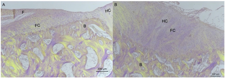
Figure 6.
Representative images of HE-stained specimens viewed with polarized light from defect treated with BMS + MSC-EVs (A) and BMS + PBS (B). On the left, bone is seen above the projected tidemark from the adjacent hyaline cartilage, while a small amount of fibrous tissue is observed superficially. On the right, a small area is seen on the right with round cells in lacunae and no collagen fibers and was scored as hyaline cartilage. The different tissue types are presented: HC = hyaline cartilage; FC = fibrocartilage; F = fibrous tissue; B = bone. Magnification: 40×, scale bar: 200 μm.

