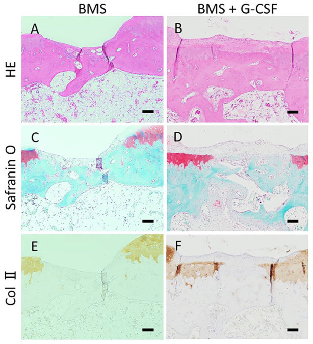
Figure 8.

Histological images in high magnification at 16 weeks. The BMS hole in Figure 7 is magnified higher. BMS holes were incompletely filled with bony structure resulting in indented surface in BMS group (A, C, E) whereas filled with bony structure nearly reaching to the original surface above with accompanying fibrous tissue in BMS + G-CSF group (B, D, F). (HE staining: A, B. Safranin-O staining: C, D. Collagen type II staining: E, F. Scale bars = 100 µm. HE, hematoxylin and eosin; Col II, collagen type II; BMS, bone marrow stimulation; G-CSF, granulocyte colony-stimulating factor).
