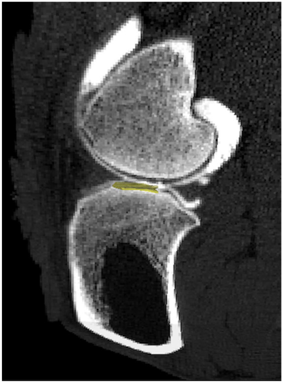
Figure 1.

Computed tomography (CT) images of contrast-enhanced sheep stifle joint in vivo. Clinical resolution CT images after a 10-minute delay following intra-articular injection of 60% anionic contrast media. Three-dimensional region of interest (ROI: 20.0mm3) (yellow) positioned within the medial tibial plateau (MTP) for apparent density analysis in cartilage in sagittal view.
