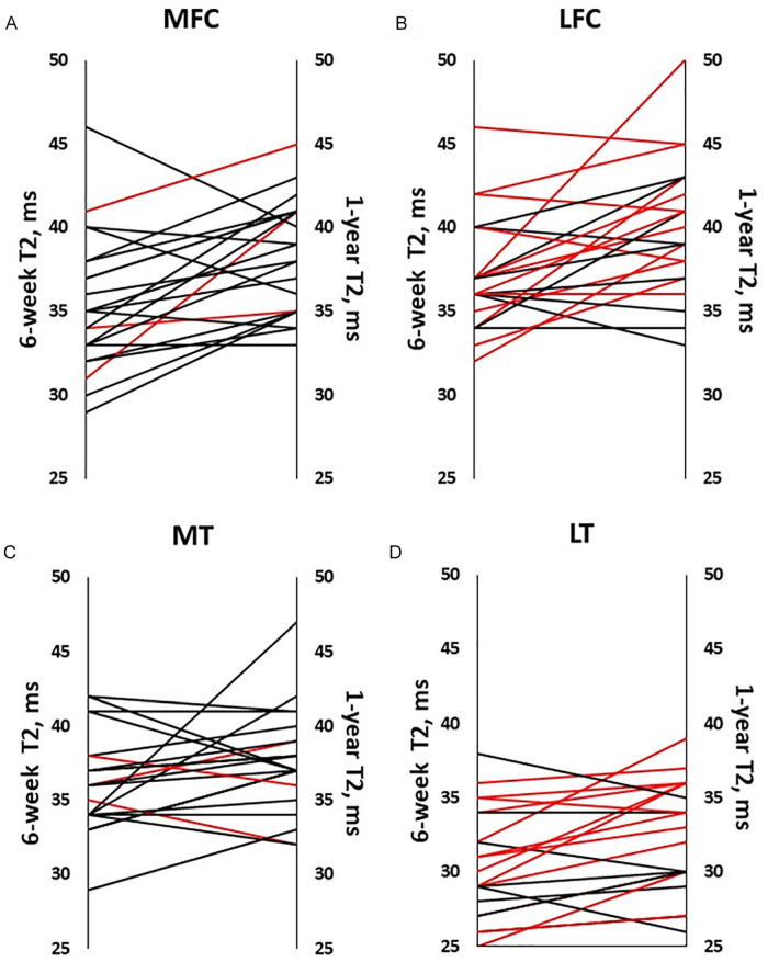
Figure 3.
Individuals’ T2 values at 6 weeks and 1 year following ACLR. T2 value averaged across all participants with ACLR increased between 6 weeks and 1 year following surgery in (A) the MFC, P = .001; (B) LFC, P = .004; (D) LT, P = .011, but not in the (C) MT, P = .130. Red lines indicate meniscal tears of the same (medial or lateral) compartment. In LT cartilage, (D) T2 increased more in participants with torn menisci treated by repair or resection than in participants with intact lateral menisci (P = .003, .046). ACLR = anterior cruciate ligament reconstruction; MFC = medial femoral condyle; LFC = lateral femoral condyle; LT = lateral tibial; MT = medial tibial.

