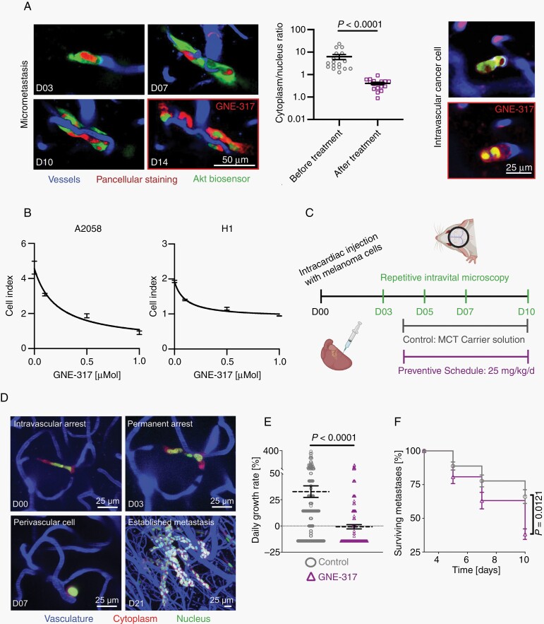
Fig. 4.
Impact of a dual PI3K/mTOR inhibitor on melanoma cells in vitro and in vivo. (A) Representative in vivo molecular imaging examples of biosensor labeled A2058 cells, showing a reduction of Akt activity in micrometastasis and intravascular arrested cancer cells in response to administration of GNE-317. Image quantification shows lower cytoplasm/nucleus ratios after administration of GNE-317. Intravital multiphoton microscopy, A2058 cells, n = 3 mice, error bars show SEM, Mann-Whitney test. (B) Real-time proliferation assays. Dose-response curves of GNE-317 treated A2058 and H1 human melanoma cells. Error bars show SD. (C) In vivo treatment study with an intravital microscopy approach investigating the response to early dual PI3K/mTOR inhibition. Mice in this preventive schedule group were treated with 25 mg/kg GNE-317 daily, control group with carrier solution. (D) Intravital multiphoton microscopy visualizes the brain metastatic cascade. A2058 cells, maximum intensity projection. (E) Tumor growth rates per day were calculated by the difference between the number of cells in metastases at day 3 and day 10. A2058 cells, n = 174/153 cells in n = 3 mice per group, data shows mean and SEM. Mann-Whitney test. (F) Metastases survival until day 10 was investigated. Proportion of surviving metastases was calculated by numbers of remaining tumor cells in relation to initially labeled cells at day 3. A2058 cells, n = 174/153 cells in n = 3 mice per group, data shows mean and SEM, Student’s t test.

