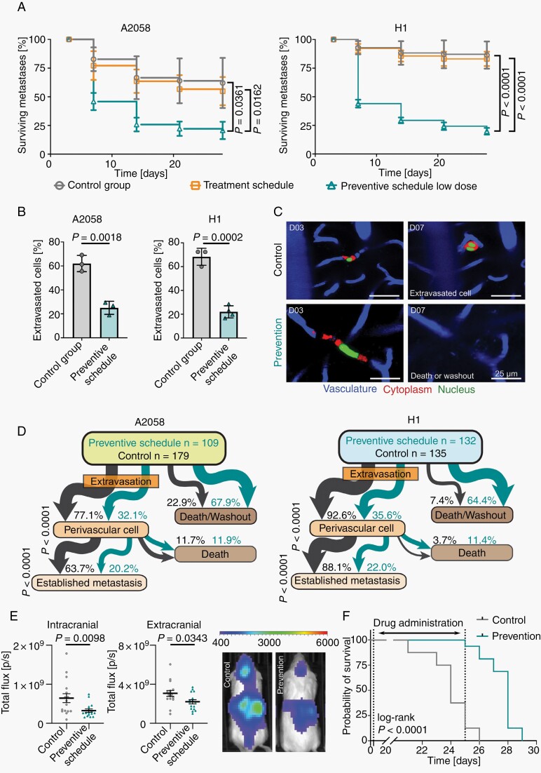
Fig. 6.
The earliest steps of the brain metastatic cascade are most vulnerable to PAM pathway inhibition. (A) Tumor cell survival between days 3 and 28 after intracardiac injection. A2058 cells, intravital multiphoton microscopy, n = 179/175/205/109 cells in n = 3 mice per group, error bars show SD, Student’s t test. H1 melanoma cells, intravital multiphoton microscopy, n = 135/77/132 cells in 3/3/4 mice per group, error bars show SD, Student’s t test. (B) Proportion of cells that performed extravasation until day 7. A2058 cells, intravital multiphoton microscopy, n = 179/175/205/109 cells in n = 3 mice per group, error bars show SD, Student’s t test. H1 cells, intravital multiphoton microscopy, n = 135/77/132 cells in 3/3/4 mice per group, error bars show SD, Student’s t test (C) Representative intravital multiphoton microscopy images show brain colonization in control group and death or wash out of a tumor cell in preventive schedule group. A2058 cells. (D) Tumor cells were followed over time, and events quantified. Flow chart indicates the fate of every individual melanoma cell from day 3 to day 28. A2058 cells, intravital multiphoton microscopy, n = 179/175/205/109, Fisher’s exact test. H1 cells, intravital multiphoton microscopy, n = 135/77/132 cells in 3/4 mice per group, Fisher’s exact test. (E) Low-dose GNE-317 administered in a preventive schedule reduced formation of intracranial and extracranial metastases of A2058 melanoma cells. Whole-body imaging (IVIS) in week 3 following intracardiac injection, n = 16 mice per group, error bars show SEM, Mann-Whitney test. (F) Mice receiving low-dose GNE-317 in a preventive schedule from day 4 to day 25 showed increased overall survival. n = 16 mice per group, log-rank test.

