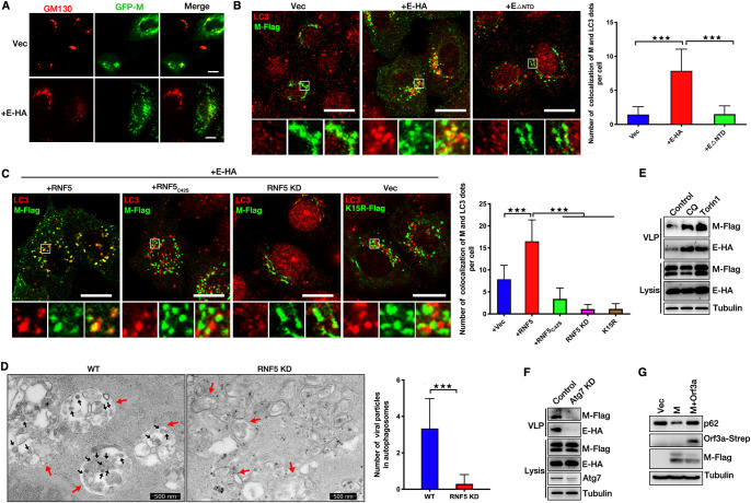
FIG 6.
RNF5 promotes the trafficking of SARS-CoV-2 M from the Golgi apparatus to autophagosomes for virions release. (A) HeLa cells were transfected with GFP-M with or without E-HA for 24 h. Cells were analyzed via immunofluorescence. GM130 antibody was used for tracking the Golgi apparatus. Scale bar, 10 μm. (B) HeLa cells were transfected with M-Flag and E-HA or EΔNTD for 24 h, and cells were analyzed via immunofluorescence. Flag antibody was used for tracking M, and LC3 antibody was used for tracking autophagosome. Scale bar, 10 μm. The graphs show the quantification of numbers of cells colocalizing with M and LC3; the numbers of dots per cell were determined by taking the average number of dots in 50 cells. Error bars, means ± SD from three experiments. Student's t test; ***, P < 0.001. (C) HeLa cells were transfected with M-Flag and E-HA with or without wild-type or C42S mutant RNF5 for 24 h or siRNF5 for 48 h, and cells were analyzed via immunofluorescence. Flag antibody was used for tracking M, and LC3 antibody was used for tracking autophagosome. Scale bar, 10 μm. The graphs show the quantification of numbers of cells colocalizing with M and LC3; the numbers of the indicated dots per cell were determined by taking the average number of dots in 50 cells. Error bars, means ± SD from three experiments. Student's t test; ***, P < 0.001. (D) Electron micrograph analysis indicates that knockdown of RNF5 abolished the accumulation of virion particles in autophagosomes. Vero cells were infected with SARS-CoV-2 for 36 h and analyzed via TEM. The black arrows indicate virion particles, and red arrows indicate autophagosomes. The graphs show the quantification of numbers of virion particles and autophagosomes that colocalized; numbers were determined by counting the average number of virion particles in 50 autophagosomes. Error bars, means ± SD from three experiments. Student's t test; ***, P < 0.001. (E) HEK293T cells were transfected with E-HA and M-Flag for 30 h. Cells were further treated with CQ or Torin1 for another 6 h. Lysates and the corresponding purified VLPs were analyzed via WB. (F) HEK293T cells were transfected with E-HA and M-Flag with or without Atg7 siRNA for 36 h. Lysates and the corresponding purified VLPs were analyzed via WB. (G) HEK293T cells were transfected with M-Flag with or without Orf3a-Strep for 36 h, and cell lysis was analyzed via WB.

