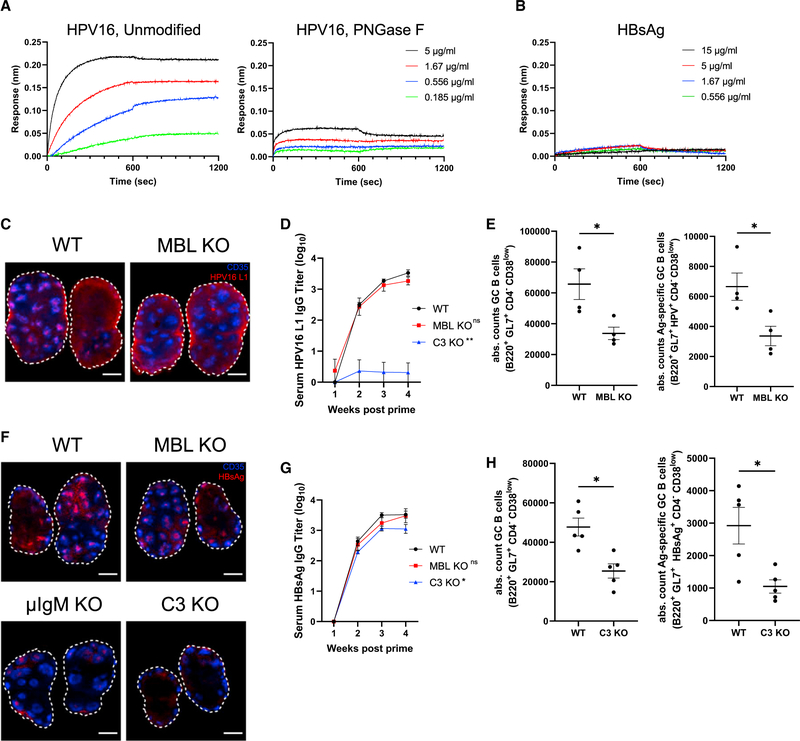
Figure 5. HPV16 L1 and HBsAg nanoparticles exhibit complement-dependent follicular accumulation and immunogenicity.
(A and B) BLI binding curves of unmodified and PNGase F-treated HPV16 L1 (A) or unmodified HBsAg (B) to immobilized recombinant murine MBL2 as functions of antigen concentration.
(C) C57Bl/6 mice or MBL KO mice (n = 5/group) were immunized with 0.1 μg AlexaFluor 647-labeled HPV16 L1 and saponin adjuvant. Seven days later, lymph nodes were harvested, cleared, and imaged by confocal microscopy. Shown are average intensity Z projections through 360 μm of tissue; shown is staining for CD35 (blue) and antigen (red), scale bars denote 500 μm.
(D) Serum HPV16 L1-specific IgG titers over time in mice (n = 5/group) immunized with 0.1 μg HPV16 L1 and saponin adjuvant. Error bars indicate SEM, p = 0.92 compared with WT one-way ANOVA.
(E) Absolute counts of germinal center B cells (B220+GL7+CD4−CD38low) and antigen-specific germinal center B cells (B220+GL7+HPV16 L1+CD4−CD38low) from WT and MBL KO mice (n = 5/group) at day 12 following immunization with 0.1 μg HPV16 L1 and saponin adjuvant. Error bars indicate SEM; *, p < 0.05 by Mann-Whitney test.
(F) C57BL/6 mice or MBL KO mice (n = 5/group) were immunized with 5 μg AlexaFluor 647-labeled HBsAg and saponin adjuvant. Seven days later, lymph nodes were harvested, cleared, and imaged by confocal microscopy. Shown are average intensity Z projections through 360 μm of tissue; shown is staining for CD35 (blue) and antigen (red), scale bars denote 500 μm.
(G) Serum HBsAg-specific IgG titers over time in mice immunized with 5 μg HBsAg and saponin adjuvant. Error bars indicate SEM; *, p <0.05 compared with WT by one-way ANOVA followed by Tukey post hoc test.
(H) Absolute counts of germinal center B cells (B220+GL7+CD4−CD38low) and antigen-specific germinal center B cells (B220+GL7+HBsAg+CD4−CD38low) from WT and C3 KO mice 12 days after immunization with 5 μg HBsAg and saponin adjuvant. Error bars indicate SEM; *, p <0.05 by Mann-Whitney test.

